1 2 3 4 5 6 7 8 9 10 11 12 13 14 15 16 17 18 19 20 21 22 23
Bacteriological analysis and antibiotic resistance in patients with suspected septicemia in Dhaka, Bangladesh (2025)
1
Bacteriological analysis and antibiotic resistance in patients with suspected
septicemia in Dhaka, Bangladesh
By
Afsana Ahmed Sohana
ID: 20226001
Mahanaz Mum Tarin Spriha
ID: 20326015
Fahmida Ahmed
ID: 21326039
A thesis submitted to the Department of Mathematics & Natural Sciences in partial fulfillment of
the requirements for the degree of
Bachelor of Science in Microbiology
Department of Mathematics and Natural Sciences,
Brac University
May 2025
2
Declaration
It is hereby declared that
1. The thesis submitted titled “Bacteriological analysis and antibiotic resistance in patients
with suspected septicemia in Dhaka, Bangladesh” is our own original work while
completing a degree at Brac University.
2. The thesis does not contain material previously published or written by a third party, except
where this is appropriately cited through full and accurate referencing.
3. The thesis does not contain material which has been accepted, or submitted, for any other
degree or diploma at a university or other institution.
4. We have acknowledged all main sources of help.
Student’s Full Name & Signature:
Afsana Ahmed Sohana
Fahmida Ahmed
20226001
21326039
Mahanaz Mum Tarin Spriha
20326015
3
Approval
The thesis titled “Bacteriological analysis and antibiotic resistance in patients with
suspected septicemia in Dhaka, Bangladesh” submitted by
1. Afsana Ahmed Sohana (20226001)
2. Fahmida Ahmed (21326039)
3. Mahanaz Mum Tarin Spriha (20326015)
In Spring, 2025 has been accepted as satisfactory in partial fulfillment of the requirement for
the degree of Bachelor of Science in Microbiology in Fall, 2024
Examining Committee:
_______________________________
Supervisor:
Fahim Kabir Monjurul Haque, PhD
(Member)
Associate Professor of Microbiology Program &
Department of Mathematics and Natural Sciences
Brac University
_______________________________
Program Coordinator:
Nadia Sultana Deen, PhD
(Member)
Associate Professor of Microbiology Program &
Department of Mathematics and Natural Sciences
Brac University
Departmental Head:
_______________________________
(Chair)
Md. Firoze H. Haque, PhD
Associate Professor and Chairperson,
Department of Mathematics and Natural Sciences
Brac University
4
Ethics Statement
This study was conducted in strict accordance with the highest ethical standards in research. Blood
samples were collected from patients at four branches of Islamic Bank Hospital in Dhaka,
Bangladesh, with full adherence to established ethical guidelines. Ethical approval was sought and
granted from the review boards of all four hospital branches prior to the initiation of the study.
These institutional review boards meticulously reviewed and approved the research protocols to
ensure compliance with the National Guidelines on Health Research and the International
Guidelines for Ethical Considerations in Research.
Furthermore, the study underwent review and received approval from the Ethics and Research
Review Committee of the Faculty of Mathematics and Natural Sciences, MNS, at BRAC
University, Dhaka, Bangladesh. The research was conducted under the supervision of Dr. Fahim
Kabir Monjurul Haque, with all aspects of the study aligning with the ethical guidelines set forth
by BRAC University.
Informed consent was obtained from the patients prior to the collection of samples. patients were
thoroughly informed about the study's objectives, the procedures involved, potential risks, and the
benefits. Data confidentiality and patient’s anonymity were rigorously maintained, with all
personal identifiers being removed during data analysis.
This robust ethical framework underpins the study's integrity and validity, ensuring adherence to
internationally recognized norms and principles for ethical research.
5
Acknowledgement
We would like to begin by expressing our deepest gratitude to Almighty Allah for granting us the
opportunity to undertake and complete this research. Our heartfelt thanks go to our parents whose
unwavering support has been invaluable from the inception to the completion of this study.
We are also deeply grateful to the Chairperson of the Department of Mathematics and Natural
Sciences, Md. Firoze H. Haque, and to all the faculty members of BRAC University for their
continuous support. Special thanks to our supervisor, Dr. Fahim Kabir Monjurul Haque,
Associate Professor in the Microbiology Program, Department of Mathematics and Natural
Sciences, BRAC University. His expert guidance, constructive feedback, and unwavering support
were crucial to the success of this research. This work would not have reached its full potential
without his dedicated mentorship and insightful critiques.
Lastly, I want to show my heartfelt appreciation to my mentor, Fahim Faisal Nakib, for his
constant clear direction and guidance. His guidance helped me ease up and navigate the journey
while enlightening and enriching the experience.
6
Table of Contents
Declaration
2
Approval
3
Ethics Statement:
4
Acknowledgment
5
Table of Contents
6
Abstract
8
List of Tables:
9
List of Figures:
10
List of acronyms:
11
CHAPTER 1
12
1.1 Introduction
12
1.2 Objectives
13
CHAPTER 2
14
Materials and Methods:
14
2.1: Study design:
14
2.2: Sample collection:
14
2.3: Culture to isolate the bacteria
14
2.4: Biochemical tests
16
2.5: DNA Extraction:
19
7
2.6: PCR (Polymerase Chain Reaction)
20
2.7: Agarose Gel Electrophoresis:
22
2.8: Antibiotic susceptibility testing:
24
CHAPTER 3
27
Result
27
3.1: Demographic characteristics of patients
27
3.2: Prevalence of positive isolated in septicemia patients
28
3.3: Antibiotic resistance & sensitive profile of the positive isolated organisms
29
3.4: Multi-drug resistant bacteria……………………………………………………………...32
CHAPTER 4
33
Discussion:
33
CHAPTER 5:
36
Conclusion:
36
CHAPTER 6
37
References:
37
8
Abstract
The objective of our study was to isolate bacteria from septicemia patient’s blood and subsequently
analyze their antibiotic resistance pattern. We collected 80 blood samples from four different
branches of Islami Bank Hospital (Mirpur, Kakrail, Motijheel, and Nayapaltan) in Dhaka,
Bangladesh. Isolating one bacterial colony from a blood culture plate of selective media. Then,
based on colony morphology, selecting the bacterial colonies and subculturing them for
presumptive identification. Confirming their identity through biochemical tests (catalase test,
oxidase test) and molecular test (polymerase chain reaction) followed by gel electrophoresis (for
visualizing the band size of targeted microorganisms). Among the 80 samples, 66 samples were
culture positive. From positive cultures, 65.2% were Salmonella typhi, 13.6% were Klebsiella
pneumoniae, 12.1% were Escherichia coli, and 9.1% were Staphylococcus aureus. 22% were
gram-positive and 77% were gram-negative) For antibiotic susceptibility tests (AST), we selected
one isolate from each culture-positive sample. Total 8 Escherichia coli, 43 Salmonella typhi, 9
Klebsiella pneumoniae, and 6 Staphylococcus aureus isolates were selected and AST was
performed using the Kirby-Bauer disc diffusion method. We observed that 90% to 100% of the
Salmonella typhi isolates 89% and 79% were resistant to Nalidixic acid and Ampicillin
respectively. 68%, 79% and 39 % of Klebsiella pneumoniae isolates were resistant to Amoxicillin,
Cefixime and Erythromycin respectively. 69% and 80% of all Escherichia coli isolates were
resistant to Amoxicillin and Cefixime respectively, and 28% of the isolates were resistant to
Azithromycin, Ciprofloxacin, Imipenem, Ceftriaxone. 49% of all Staphylococcus aureus isolates
were resistant to Cefixime, Sulfamethoxazole and Ceftazidime,
28% were resistant to
Erythromycin and Ceftriaxone, and 19% were resistant to Imipenem and Gentamicin. In this study,
a total of 18 isolates were identified as multi-drug resistant (MDR), including 7 Salmonella typhi,
6 Klebsiella pneumoniae, 2 Escherichia coli, and 3 Staphylococcus aureus strains. Among these,
3 isolates were further classified as extensively drug-resistant (XDR), comprising 1 Salmonella
typhi, 1 Klebsiella pneumoniae, and 1 Staphylococcus aureus isolate.
Keywords: Septicemia, Salmonella typhi, Klebsiella pneumoniae, Escherichia coli,
Staphylococcus aureus, Multi-drug resistant (MDR), Extensively drug-resistant (XDR).
9
List of Tables:
Table No.
Table Title
Page No.
1.
Desired colony morphology of isolated bacteria
15
2.
Biochemical test results of the organism
18
3.
List of targeted genes and of their primers
21
4.
Concentrations and interpretive criteria for
26
diffusion zones of the antibiotics.
5.
Demographic characteristics of patients
27
10
List of Figures:
Figure No.
Figure Title
Page No.
1.
Different bacterial colonies of different selective media
16
2.4
Different type of biochemical tests
2(a).
Agarose gel electrophoresis of PCR assay of Klebsiella
22
pneumoniae isolates
2(b).
Agarose gel electrophoresis of PCR assay of Staphylococcus
23
aureus isolates
2(c).
Agarose gel electrophoresis of PCR assay of Salmonella typhi
23
isolates
2(d).
Agarose gel electrophoresis of PCR assay of Escherichia coli
24
isolates
3.
Prevalence of Isolated pathogens in Septicemia Patients based
28
on positive results
4(a).
Antibiotic Susceptibility Profiling of Salmonella typhi
29
positive isolates
4(b).
Antibiotic Susceptibility Profiling of Klebsiella pneumoniae
30
positive isolates
4(c).
Antibiotic Susceptibility Profiling of Escherichia coli
31
positive isolates
4(d).
Antibiotic Susceptibility Profiling of
32
Staphylococcus aureus positive isolates
11
List of Acronyms
PCR
Polymerase Chain Reaction
AST
Antibiotic Susceptibility testing
MHA
Muller-Hinton Agar
MDR
Multi Drug Resistant
XDR
Extensive Drug Resistant
XLD
Xylose Lysine Deoxycholate
EMB
Eosin Methylene Blue
MAC
MacConkey Agar
MSA
Mannitol Salt Agar
12
CHAPTER 1
1.1: Introduction
Sepsis is a hazardous organ dysfunction caused by an immune response to infection. And so it is
a leading cause of death, especially in low income countries like Bangladesh. In developing
countries like Bangladesh, India or Pakistan, the leading causes of infection are malaria,
pneumonia and diarrhea. Here, sepsis dominates because of higher malnutrition, lack of certified
doctors, poor healthcare infrastructure, and late sepsis detection. If left untreated, sepsis can
manifest in more severe forms like septic shock, multiple organ failure, and finally death. That’s
why it is important to detect sepsis as soon as possible. We also need more information to design
the most appropriate treatment method for each sepsis case. (Garbern et al., 2024) At present,
special colorimetric assays can detect septicemia at early stages, by tracking special biomarkers in
blood. This is a very important discovery for medical science. Because a patient’s life depends on
the earliest possible availability to treatment. (Dashtian et al., 2024). This has been confirmed in a
review of 42 studies on 19000 patients and more. All the findings confirm that the death risks of
septicemia drop by 16 percent if the patients receive treatment within the first 3 hours of diagnosis.
After this time has passed, the chances of death increase by 30 per cent. The results are published
in the European Journal of International Medicine (Leung et al., 2024). But sadly, antibiotic
resistance is on the rise (confirmed by the World Health Organization). This is a huge obstacle to
treatment (Baral et al., 2024). And most of the causative agents of septicemic pathogens are already
resistant to the commonly used antibiotics. For example, a study in Turkey shows that deaths of
48% of infected patients are caused by gram negative sepsis. Infections from Methicillin resistant
Staphylococcus aureus cause 36 % of deaths. Carbapenem-resistant pathogens can kill 60 percent
of patients on average. (Gucer et al., 2024). Another research in Ethiopia showed that 89 percent
of nosocomial pathogens are already resistant to multiple drugs (like ampicillin or amoxicillin.)
Among them, 34.7% are resistant to carbapenems and 46.3 percent to beta lactams (Tilahun et al.,
2024). These statistics are concerning. So, these pathogens should be studied extensively. And
appropriate antibiotics should be designed to stop their growth, so that antibiotic resistance can be
slowed down or reversed, if possible. (Baral et al., 2024). Even Bangladesh is trying to design
proper treatment plans for septicemia. Experts from other countries are also trying to find solutions
in our country, like Dr. Stephanie Garbern from Brown University, United States. She mainly
13
studied children affected with septicemia in poor and middle-income countries. She even worked
with engineers and local doctors to develop an effective diagnostic gadget. It was also trained with
a machine learning algorithm, which can collect the person’s data (heart rate, oxygen rates etc.)
Then the AI can detect early symptoms of sepsis, which doctors might mistake as fever or common
cold. She only used cheap, available tools so that poor and middle-class people can access it too.
(Garbern et al., 2024). Nevertheless, it is important to find cost effective ways to diagnose and
treat septicemia in a third world country like Bangladesh.
Despite the discovery of advanced treatment methods for septicemia, antibiotics are the standard
treatment plan because it directly targets the pathogens, who are the main agents of disease. That’s
why we felt the need to conduct this research- to find the ideal antibiotic to kill each pathogen. But
antibiotics are losing their potency because of some malpractices in our Healthcare system.
Antibiotic usage is largely unregulated in Bangladesh, especially in rural areas. Without diagnosis,
pharmacists and doctors prescribe multiple antibiotics to the customers without diagnosis. These
antibiotic sellers don't know the guidelines of using antibiotics. In addition, patients don't complete
their medication for the full prescribed time, which buys time for bacteria to develop antibiotic
resistant genes (Bepari et al., 2023). Extensive studies have been conducted on this pattern of
emergence. A review article analyses 46 studies from 2014 to 2018, which reveals high prevalence
of antibiotic resistance among common pathogens. (Ahmed et al., 2019).
Regarding the growing resistance to antibiotics, it is essential to conduct regular studies to identify
the bacteria causing septicemia among Bangladeshi patients and assess their antibiotic resistance.
This helps to monitor the changes in antibiotic resistance. Additionally, this will provide doctors
and authorities with insights to develop treatment strategies that prescribe efficient antibiotics for
septicemia patients.
1.2: Objectives
The objectives of this study were to isolate the bacteria causing septicemia from septicemia-
suspected patients in Dhaka city and to analyze their antibiotic resistance patterns.
14
CHAPTER 2
Materials and Methods
2.1 Study design
The study was conducted among clinically suspected septicemia patients who were admitted to
hospitals in Dhaka, Bangladesh. Samples were collected from four branches of Islami Bank
Hospitals; Mirpur, Motijheel, Kakrail and Naya paltan & 80 samples were collected from these
branches. The blood sample collection duration was from December,2024 to April, 2025. Blood
samples were collected from patients of various age groups and both genders who were admitted
to various wards including the Intensive Care Unit (ICU), Medicine, Pediatrics, and Emergency
wards. Written consent was taken prior to sample collection, from either patients or the legal
guardians of patients.
2.2 Sample collection
Venous blood samples were taken from clinically confirmed septicemia patients upon admission
to hospitals that were chosen for the study. Venipuncture was used to obtain blood samples using
sterilized, disposable syringes and needles by professional experts. Before beginning antibiotic
treatment, each patient had a blood draw of about 2-3 mL. Blood was immediately put into a
heparin tube which was transported to the microbiology lab via a sealed iced box(4°C). The
samples were appropriately labeled with the information of patient's ID, date, name, gender and
indoor/outdoor patients. After being collected, they were taken to the microbiological lab in 1-2
hours(Nursing et al., 2023).
2.3 Culture to isolate the bacteria
Upon arrival at the laboratory, each blood sample was immediately processed to ensure the
integrity of microbial content. A volume of 100 µL of the blood sample was aseptically dispensed
and spread onto various culture media. This was carried out using a sterile micropipette and
spreader under laminar flow conditions to minimize contamination. The selected media types
15
included MacConkey (MAC), Xylose Lysine Deoxycholate (XLD), Eosin Methylene Blue
(EMB), Mannitol Salt Agar (MSA), HiCrome™ UTI, HiCrome™ KPC Agar media. They were
then incubated at 37°C and checked 5-7 days for indications of microbial growth such as changes
in medium color and colonies. After up to seven days of incubation, spread plates were observed
if there is any growth. Positive cultures that showed evidence of growth were sub cultured onto
selective and differential media such as Salmonella typhi on SS Agar media, XLD medium,
Escherichia coli on EMB agar, HiCrome™ UTI Agar, klebsiella pneumoniae on KPC,
Staphylococcus aureus on Mannitol Salt Agar. Thus, expected colony morphology of these
organisms has shown in Table 1. For 18 to 24 hours, plates were incubated at 37°C, and the visible
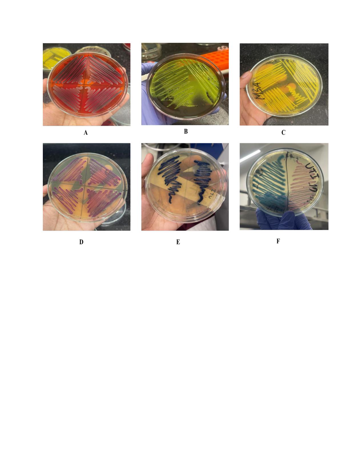
of bacterial colonies was observed and has shown in the Figure 1.
Table 1: Desired colony morphology of isolated bacteria
Organism
Gram
Media
Expected Colony
Positive/Negative
Morphology
Staphylococcus aureus
Gram-positive
Yellow/white colonies
Mannitol Salt Agar
surrounded by the yellow
zone
Klebsiella pneumoniae
Gram-negative
HiCrome UTI Agar
Blue, mucoid
Klebsiella pneumoniae
Gram-negative
HiCrome KPC Agar
Bluish-green
Escherichia coli
Gram-negative
HiCrome UTI Agar
Purple colonies
Escherichia coli
Gram-negative
EMB Agar
Purple with a black center
and green metallic sheen
Salmonella Shigella
Salmonella typhi
Gram-negative
Agar
Colorless with black centered
Xylose-Lysine-
Salmonella typhi
Gram-negative
Red with black centered
Deoxycholate agar
16
Figure 1 : Different bacterial colonies of different selective media. (A) Salmonella typhi has given
black centered on XLD agar, (B) Escherichia coli shows green metallic sheen on EMB agar, (C)
Staphylococcus aureus gives yellow colored colony on MSA, (D) Escherichia coli has given
purple colony on HiCrome UTI Agar, (E) Klebsiella pneumoniae has given blue colony on
HiCrome KPC Agar & lastly (F) Escherichia coli with purple colony & Klebsiella pneumoniae
with blue colony which distinguish them on UTI media.
2.4 Biochemical tests
Biochemical tests were performed to analyze the metabolic and enzymatic properties of the
isolated organisms. Here, catalase, oxidase tests were done for Staphylococcus aureus, Escherichia
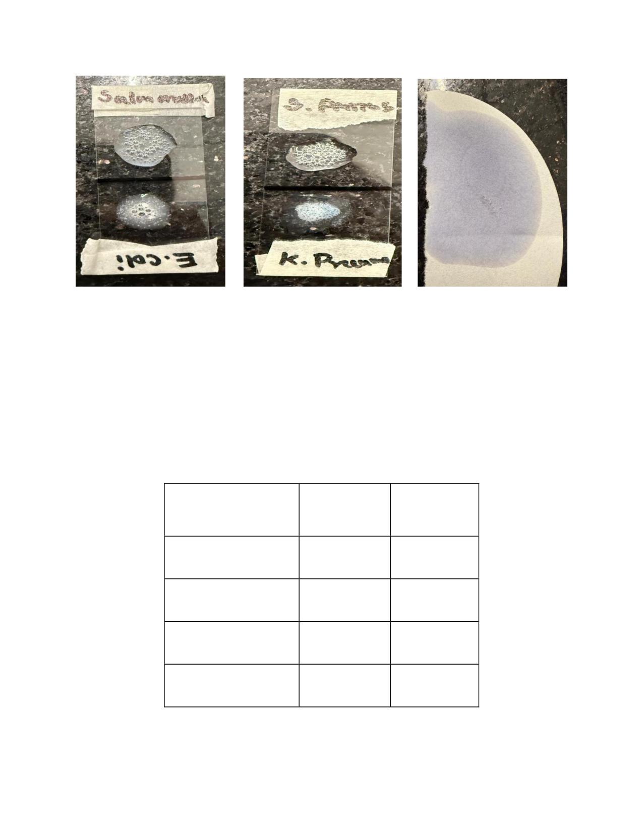
coli, Salmonella typhi & Klebsiella pneumoniae. Table 2 shows the biochemical test results for
the isolated bacteria.
17
Catalase test method:
1)Spread the bacteria on an agar plate and incubate it overnight (18-24 hours) under the proper
conditions.
2)Using a sterile inoculating loop, select bacteria from a single colony and transfer them to a
microscope slide.
3)After adding one drop of 3% H2O2 to the bacteria, the suspension will give the result whether
it is positive or negative.
● Positive test result: Gas formation (O2) in the form of bubbles shows that the bacterium
has a catalase.
● Negative test result: No Gas formation (O2) in the form of bubbles.
Oxidase test method:
1)On a piece of filter paper, apply two drops of the oxidase reagent.
2)Using a sterile loop to transfer bacteria from one colony onto the oxidase reagent area. The result
can be observed after 30 sec.
● Positive test result: Dark blue-purple color change within 10-30 sec.
● Negative test result: No color change or color change after more than 30 sec (VETBact,
n.d).
18
(A)
(B)
(C)
Figure 2.4: Positive results of catalase tests of Salmonella typhi & Escherichia coli (A),
Klebsiella pneumonia & Staphylococcus aureus (B) is shown in the above figure. Negative
results of oxidase tests of Salmonella typhi, Escherichia coli, Staphylococcus aureus &
Klebsiella pneumonoiae (C).
Table 2: Biochemical test results of the organism
Organism
Catalase
Oxidase
Staphylococcus aureus
+ve
-ve
Escherichia coli
+ve
-ve
Salmonella typhi
+ve
-ve
Klebsiella pneumoniae
+ve
-ve
19
2.5 DNA extraction
DNA was extracted for the purpose of identifying the organism by visualizing their band
production in gel electrophoresis followed by PCR. For the isolation of high-quality bacterial
genomic DNA, a freshly grown subculture was prepared using nutrient-rich growth media. As a
base solution for cell lysis, 1X Tris-EDTA buffer was prepared before the DNA extraction process
started. First, an Eppendorf tube was filled with 400 µL of 1X TE buffer. The buffer-containing
tube was then inoculated with a loopful of the bacterial culture. After that, the sample was then
vortexed to thoroughly mix the TE buffer with the bacterial cells for 15 seconds. To pellet the
bacterial cells, the sample was centrifuged for 10 minutes at 13,000 rpm and 25°C after initial
mixing. After centrifugation, the supernatant was discarded and 400 µL of 1X TE buffer was added
to the Eppendorf tube along with the pellet. The bacterial pellet was then resuspended by vortexing
the sample once more. In order to rupture the bacterial cells and liberate their genomic DNA, the
samples were to undergo a heat treatment. For ten minutes, the tubes were kept at 100°C in a heat
block machine. The bacterial cell wall and membrane will be properly lysed by this heat treatment,
releasing the DNA into the solution. Following this heat block process, the samples were allowed
to cool for five minutes at room temperature. To remove the recovered DNA from any leftover
debris or biological components, the cooled samples were centrifuged for 10 minutes at 13,000
rpm. Lastly, 200 µL of the DNA supernatant was transferred to a new Eppendorf tube after these
centrifugation procedures. Extreme caution was used to avoid disturbing the pellet at the tube's
bottom during the transfer. The isolated DNA was now present in this supernatant, making it
suitable for additional examination. Then store the DNA at -20° temperature. For use in PCR or
sequencing, this technique guaranteed the effective extraction of high-quality DNA from the
bacterial cells (Moore et al,2005).
20
2.6 PCR (Polymerase Chain Reaction)
To confirm the identity of the bacterial isolates, Polymerase Chain Reaction (PCR) was performed.
This molecular method was utilized to detect organism-specific genetic sequences.
PCR method
PCR cycle is carried out 25-40 times in order to amplify the DNA according to the organisms. To
initiate the PCR process, each reaction mixture was prepared in a final volume of 13 μL. The
reaction mixture included 3 μL of nuclease-free water, 1 μL of forward primer, 1 μL of reverse
primer, 6 μL of Emerald Amp PCR Master Mix (Takara Bio Inc.), and 2 μL of template DNA. All
components were carefully added into sterile PCR tubes. Following preparation, the tubes were
vortexed gently for 3-5 seconds to ensure thorough mixing of reagents. The mixtures were then
briefly centrifuged to collect contents at the bottom of the tubes before being loaded into a thermal
cycler (Applied Biosystems 2720 Thermal Cycler). PCR amplification was carried out under
standardized cycling conditions. Typically, 25-35 amplification cycles were performed which
involved denaturation, primer annealing, and extension steps-allowing exponential amplification
of the target DNA region. The total run time for the PCR ranged from 1.5 to 3 hours, depending
on the specific protocol and target DNA fragment length. Upon completion, all amplified PCR
products were stored at -20°C to preserve sample integrity for downstream applications such as
gel electrophoresis or sequencing (Lorenz, 2012).
Targeted genes and their primers:
This study focused on detecting virulence-associated genes specific to Escherichia coli, Klebsiella
pneumoniae, Staphylococcus aureus, and Salmonella Typhi in order to screen blood samples for
the presence of pathogenic strains, making it essential to identify the genetic markers associated
with their pathogenicity. For example, these four microorganisms are prominent etiological agents
in septicemia, each with distinct virulence factors. Table 3 shows the list of targeted genes &
primers of these organisms. This investigation aimed to detect these bacterial pathogens in blood
samples by targeting their specific virulence genes, thereby contributing to a better understanding
of the microbial causes of septicemia.

21
Table 3: List of targeted genes and of their primers
Name of
Primer
Targeted genes
Primer Sequence
Prod
PCR conditions
References
bacteria
Designati
(5′to 3′)
uct
on
size
Klebsiella
KP pf-F
16S-23S rRNA
ATTTGAAGAGGT
130
The cycling conditions were 10
(Kot et al.,
pneumoni
Internal
TGCAAACGAT
bp
min at 94°C followed by
35
2023)
ae
Transcribed
cycles of 30 s at 94°C, 20 s at
Spacer (ITS)
57°C and 20 s at 72°C, followed
by a 10 min final extension 72°C.
KP pr1-R
16S-23S rRNA
TTCACTCTGAAG
Internal
TTTTCTTGTGTTC
Transcribed
Spacer (ITS)
Escherich
ECO- F
malB (maltose
GACCTCGGTTTA
585
Initial denaturation at 95°C for 5
(Lindsey et
ia coli
operon)
GTTCACAGA
bp
min; 35 cycles of denaturation at
al., 2017)
94°C for 45s, annealing at 45°C
for 45s, and extension for 1 min
followed by a final extension at
ECO-R
malB (maltose
CACACGCTGACG
72°C for 5 min.
operon)
CTGACCA
Salmonell
Salmonell
invA
GTATTGTTGATT
403
Initial denaturation at 95°C for 5
(Ranjbar et
a typhi
a-F
AATGACATCCG
bp
min; 35 cycles of denaturation at
al., 2016)
95°C for 30s, annealing at 60°C
for 30s, and extension for 1 min
at
72°C followed by a final
Salmonell
invA
ATATTACGCTAC
extension at 72°C for 10 min.
a-R
GGAAACACGTT
Staphyloc
NUC-F
nuc
GCGAT
279
4 min at 94°C and then 34 cycles
(Gogoi et
occus
GATGGTGATACG
bp
of 1min at
94°C for
the
al., 2024)
aureus
GTT
denaturation step and
45s at
55°C for the annealing &
extension at
72°C for
45s
followed by final extension at
NUC-R
nuc
AGCCAAGCCTTG
72°C For 10min.
ACGAACTAAAGC
22
2.7 Agarose gel electrophoresis
After performing Polymerase Chain Reaction, the resulting DNA fragments were analyzed
through Agarose gel electrophoresis method. This technique was employed to visualize DNA
fragment sizes and confirm the presence of DNA by comparison with a DNA ladder. To prepare
the gel, agarose powder (typically 1.5-2%) is mixed with 2000 μL of 50x Tris-acetate-EDTA
(TAE) buffer and distilled water (98%) and heated until fully dissolved. Ethidium bromide, an
intercalating agent that fluoresces under UV light, is added at a concentration of 4 μL to enable
DNA visualization. PCR products are carefully loaded into the wells of the prepared gel using
sterilized (autoclaved) micropipette tips. The gel is run using a 10x TAE buffer as the running
buffer, facilitating the movement of DNA toward the positive pole. For each run, 4 μL of a 100 bp
DNA ladder (Biolab) is used as a reference and electrophoresis is carried out at 100 volts at 400
amp. Once the electrophoresis is complete, the gel is observed under UV light. The DNA bands
become visible due to binding of the fluorescent stain, which emits specific colors. Band sizes are
then determined by comparing them to the bands in the DNA ladder, and the results are recorded
accordingly (Addgene: Protocol - How to Run an Agarose Gel, n.d.). Below the four types of band
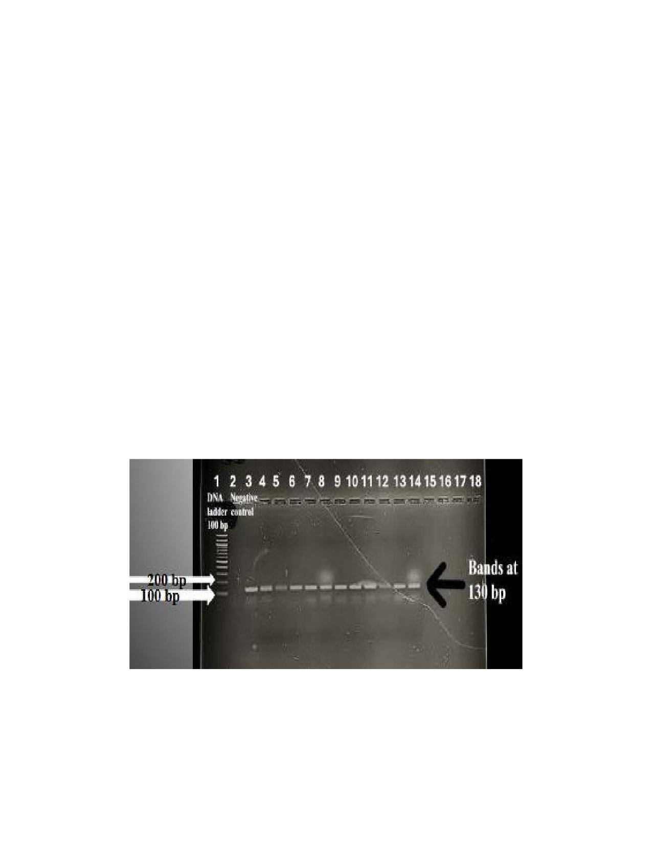
sizes have been shown in Figure 2(a), 2(b), 2(c), and 2(d).
Figure 2(a): Agarose gel electrophoresis of PCR assay of Klebsiella pneumoniae isolates. Here,
first lane is 100 bp DNA ladder, 2nd lane is negative control and well 3-14 are positive samples at
130 bp. The image was captured using a camera while the agarose gel was positioned under a UV
transilluminator. The complete original image was utilized to generate the figure panel.
23
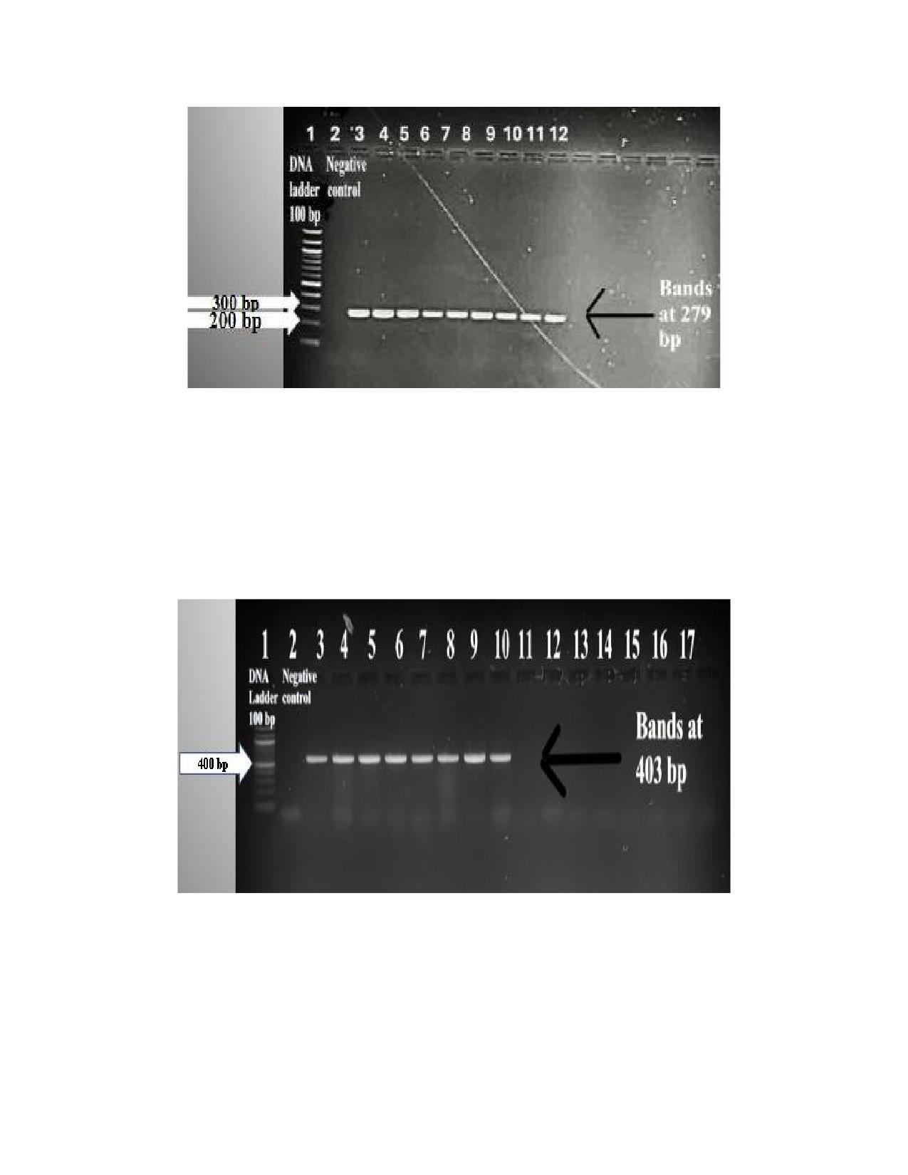
Figure 2(b): Agarose gel electrophoresis of PCR assay of Staphylococcus aureus isolates. Here,
first lane is loaded with a 100 bp DNA ladder, the 2nd lane is negative control and lanes 3-11 are
positive samples at 279 bp. The image was captured using a camera while the agarose gel was
positioned under a UV transilluminator. The complete original image was utilized to generate the
figure panel.
Figure 2(c): Agarose gel electrophoresis of PCR assay of Salmonella typhi isolates. Here, first
lane is 100 bp DNA ladder, 2nd lane is negative control and lane no. 3-10 are positive samples at
403bp. The image was captured using a camera while the agarose gel was positioned under a UV
transilluminator. The complete original image was utilized to generate the figure panel.
24
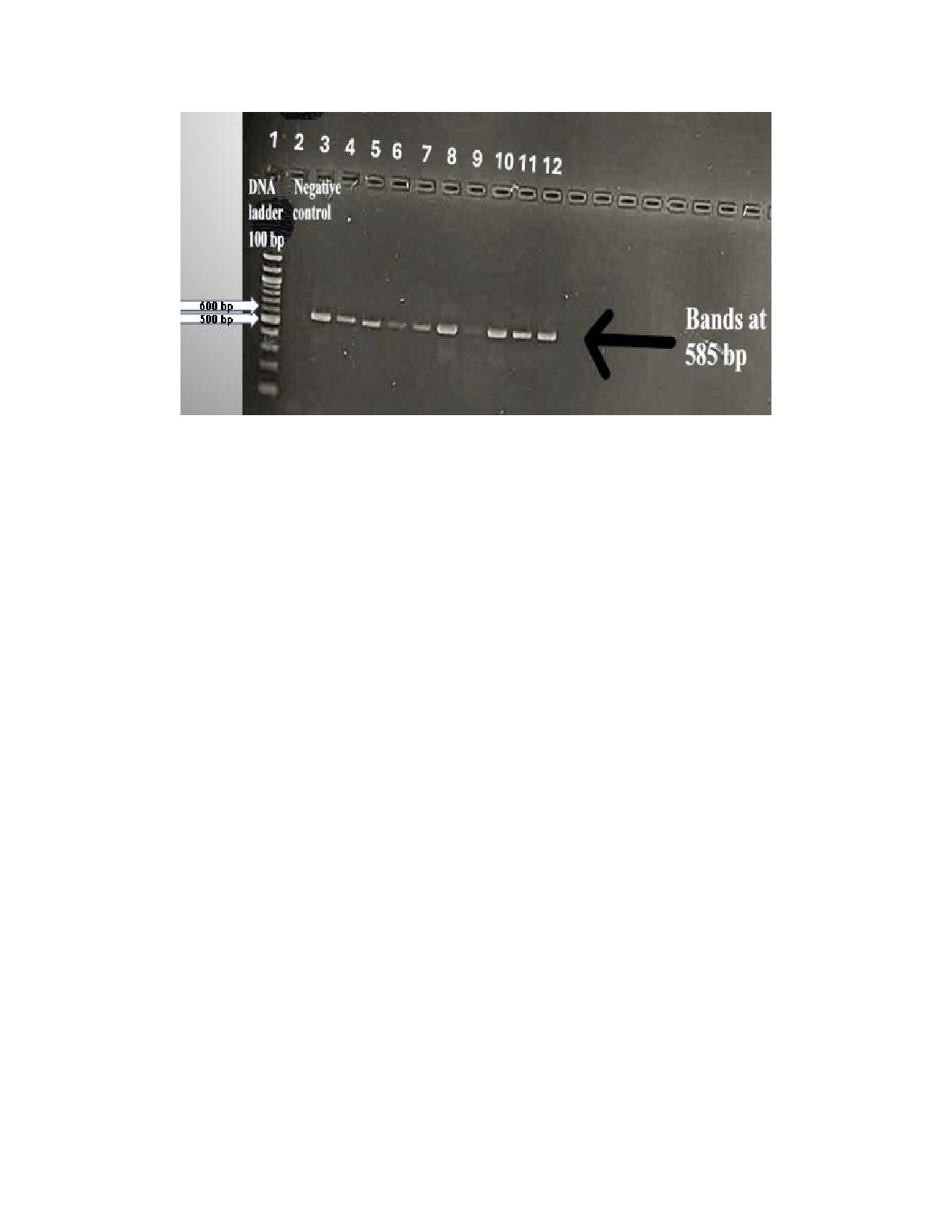
Figure 2(d): Agarose gel electrophoresis of PCR assay of Escherichia coli isolates. Here, first
lane is a 100 bp DNA ladder, the second lane is negative control and well 3-12 are positive samples
at 585 bp. The image was captured using a camera while the agarose gel was positioned under a
UV transilluminator. The complete original image was utilized to generate the figure panel.
2.8 Antibiotic susceptibility testing
The Antibiotic Susceptibility Test (AST) was performed using the Kirby-Bauer disk diffusion
method to determine the effectiveness of various antibiotics against the isolated bacteria. Each
bacterial isolate from the culture plates was sub-cultured on freshly prepared nutrient agar to obtain
a fresh and pure culture before performing AST. Mueller-Hinton Agar (MHA) media was freshly
prepared for antibiotic susceptibility testing. A 0.9% sodium chloride (NaCl) solution was also
prepared by dissolving the appropriate amount of NaCl in distilled water and briefly heating it.
Using a sterile glass pipette, 5-6 mL of the NaCl solution was dispensed into each test tube. A
bacterial suspension was prepared by picking colonies from a pure culture and adjusting the
turbidity to match the 0.5 McFarland standard. The suspension was then evenly spread over the
surface of a Mueller-Hinton agar plate using a sterile cotton swab to ensure a uniform lawn of
bacterial growth. After allowing the plate to dry for a few minutes, antibiotic discs were placed on
the agar surface using sterile forceps. The plates were incubated at 35-37°C for 16-18 hours (Jan
Hudzicki, 2009).
25
Zone Of inhibition:
Following 24 hours of incubation, the Mueller-Hinton Agar (MHA) plates were examined for
bacterial growth surrounding the antibiotic discs. If an antibiotic effectively inhibits the test
organism, a clear area devoid of bacterial growth-known as the zone of inhibition-will be
observed around the disc. In contrast, if the antibiotic is less effective or ineffective, bacterial
growth will extend closer to the disc, resulting in a smaller or no visible inhibition zone. The
diameter of each zone of inhibition is measured using a ruler or scale to accurately assess the
degree of bacterial growth suppression. These measurements are then interpreted using
standardized reference charts provided by Oxoid Limited (England), which classify the bacteria as
sensitive, intermediate, or resistant to the antibiotics tested. This interpretation method aligns with
the guidelines recommended by the National Antimicrobial Resistance Monitoring System and the
Clinical and Laboratory Standards Institute (CLSI, 1997, 1999). Table 4: shows the concentration
and interpretive criteria for diffusion zones of the antibiotics.

26
Table 4: Concentrations and interpretive criteria for diffusion zones of the antibiotics.
Antibiotic
Group
Effective against
Disc
Disc
Interpretative Criteria
code
potency
Sensitive
Intermediate
Resistant
(µg)
mm or more
mm
mm or less
Azithromycin
Macrolide
Gram-positive
and
AZM
15
18
14-17
13
Gram-negative
Amikacin
Aminoglycoside
Gram-positive
and
AK
30
17
15-16
14
Gram-negative
Colistin
Polymyxin E
Gram-negative
CL
10
-
11-17
-
Cefepime
Cephalosporin
Gram-positive
and
CPM
30
25
19-24
18
Gram-negative
Meropenem
Carbapenem
Gram-positive
and
MEM
10
23
20-22
19
Gram-negative
Imipenem
Carbapenem
Gram-positive
and
IMI
10
23
20-22
19
Gram-negative
Nalidixic Acid
Quinolone
Gram-negative
NA
30
19
14-18
13
Streptomycin
Aminoglycoside
Gram-positive
and
S
10
15
12-14
11
Gram-negative
Ampicillin
Beta-lactamase
Gram-positive
and
AMP
10
17
14-16
13
Gram-negative
Amoxicillin
Beta-lactam
Gram-positive
and
AML
10 µg
17
14-16
13
(Penicillin)
Gram-negative
Vancomycin
Glycopeptide
Gram-positive
VA
30
17
15-16
14
Linezolid
Oxazolidinones
Gram-positive
LZ
30
23
21-22
20
Tigecycline
Glycylcycline
Gram-positive
and
TGC
15
18
15-17
15
Gram-negative
Tetracycline
Tetracycline class
Broad-spectrum (Gram-
TE
30
15
12-14
11
positive and Gram-
negative)
Ciprofloxacin
Fluoroquinolone
Broad-spectrum Gram-
CIP
5
21
16-20
15
negative
Chloramphenicol
Amphenicol
Broad-spectrum (Gram-
C
30
18
13-17
12
positive and Gram-
negative)
Cefixime
3rd
Gen
Gram-negative
CFM
5
19
16-18
15
Cephalosporin
Ceftazidime
3rd
Gen
Gram-negative
CAZ
30
18
15-17
14
Cephalosporin
Erythromycin
Macrolide
Mainly Gram-positive,
E
15
23
14-22
13
some Gram-negative
cocci
Gentamicin
Aminoglycoside
Broad-spectrum;
CN
10
15
13-14
12
effective mainly against
Gram-negative
and
some
Gram-positive
bacteria
Trimethoprim-
Sulfonamide
Broad-spectrum; many
SXT
25
16
11-15
10
Sulfamethoxazole
combination
Gram-positive
and
Gram-negative bacteria

27
CHAPTER 3
Result
3.1 Demographic characteristics of patients
This study included a total of 80 participants, individuals sourced from Islami Bank Hospital from
Mirpur, Motijheel, Kakrail, Naya Paltan branches of Dhaka. Among these participants,
15(18.75%) from Islamic Bank Hospital Motijheel branch, while 40 (50%) were from Mirpur
branch & the remaining 15(18.75%) & 10(12.5%) patients from Naya Paltan branch & Kakrail
branch. The majority of the study’s subjects fell within the age range of 40 to 80 years old.
Moreover, male participants take more account for this study with a percentage of 56.25% &
remaining 43.75% is female participants.Among the patients, most of them acquired infection
through community & after that they got admitted to the hospitals. A summary of the demographic
characteristics of the study’s patients is presented in Table 5.
Table 5: Demographic characteristics of patients
Characteristics
Categories
Frequency
Percentage
Number of participants
Islamic Bank Hospital, Mirpur
40
50%
at this study
Islamic Bank Hospital, Motijheel
15
18.75%
Islamic Bank Hospital, Kakrail
10
12.5%
Islamic Bank Hospital, Nayapaltan
15
18.75%
Gender
Male
45
56.25%
Female
35
43.75%
Age(in years)
0-19
10
12.5%
20-39
15
18.75%
40-59
20
25%
60+
35
43.75%
Patient type
In patient
25
31.25%
Outpatient
55
68.75%
28
3.2 Prevalence of positive isolates in septicemia patients
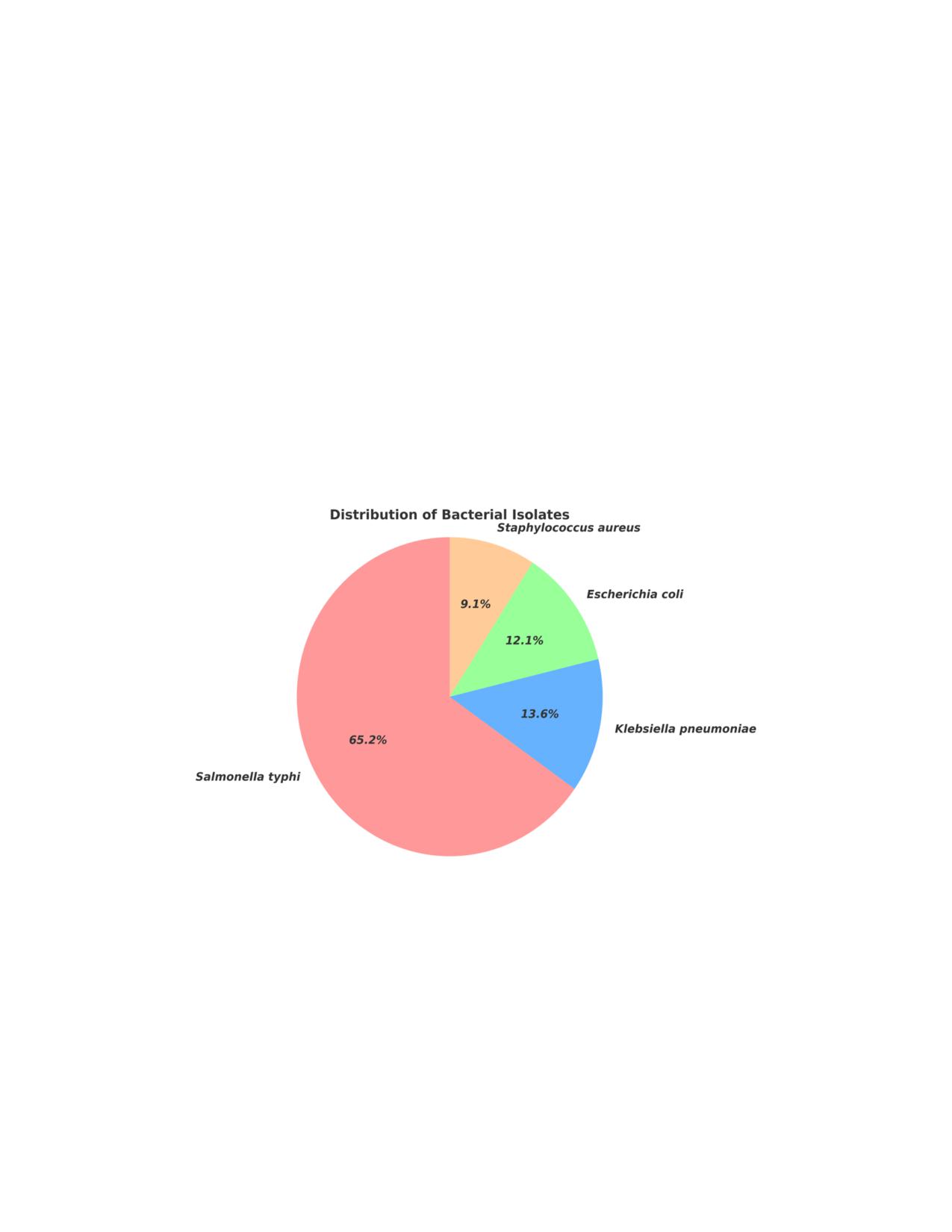
Among 80 samples, bacterial growth was observed in 66 samples. The distribution of the bacteria
in these culture-positive samples are shown in Figure 3. The data indicates that Salmonella typhi
is the most prevalent pathogen, responsible for 65.2% of the positive isolates, with 43 out of 80
samples yielding positive results. Klebsiella pneumoniae follows with 9 positive isolates,
representing 13.6% of the total, while E. coli is identified in 8 isolates, accounting for 12.1%.
Staphylococcus aureus appears in 6 isolates, making up 9.1% of the cases. All infections were due
to a single organism. These findings reflect the distribution of bacterial pathogens in septicemia
cases and emphasize the dominance of Salmonella typhi in the observed patient population.
Figure 3: Prevalence of isolated bacteria in 66 culture-positive samples of suspected septicemia
patients. This pie chart shows the distribution of isolated pathogens identified in septicemia
patients. Salmonella typhi at 65.2% of the total isolates, followed by Klebsiella pneumoniae at
13.6%, Escherichia coli at 12.1%, and Staphylococcus aureus at 9.1%.
29
3.3 Antibiotic resistance & sensitive profile of the positive isolated organisms
In this study, a total of 80 isolates have been selected to identify the antibiotic resistance of
different organisms. The Kirby-Bauer Disc Diffusion test was performed on each isolate. A total
of 8 isolates of Escherichia coli, 43 isolates of Salmonella typhi, 9 isolates of Klebsiella
pneumoniae, and 6 isolates of Staphylococcus aureus were examined for antibiotic susceptibility.
Figure 4(a), 4(b), 4(c), 4(d) demonstrates all antibiotic susceptibility profiles for specific bacteria.
Figure 5 shows the MDR, XDR & susceptible of all the isolates. In order to get the best possible
treatment results, these findings highlight the significance of concentrating on highly efficient
antibiotics while avoiding those with high resistance.
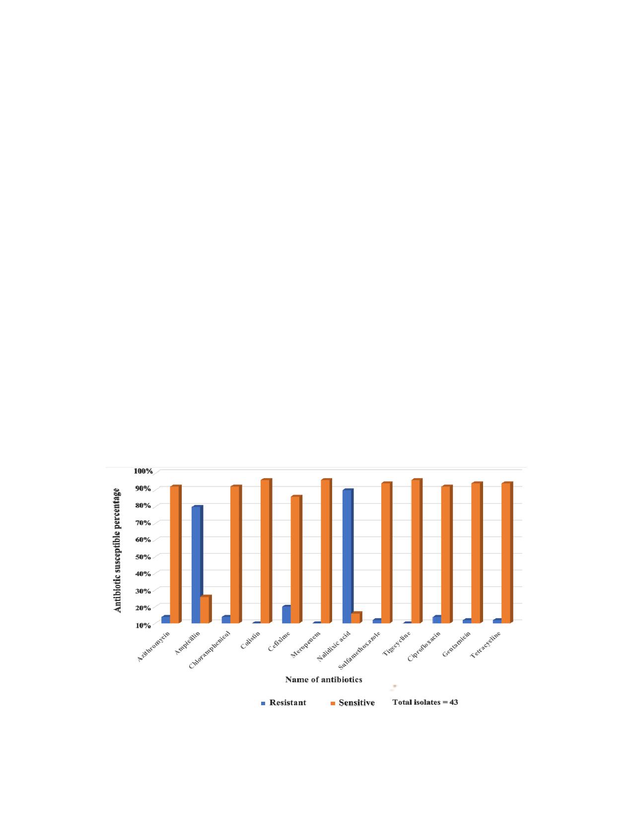
Antibiotic Susceptibility Analysis of Salmonella typhi:
Out of the all-positive Salmonella typhi isolates showed relatively high antibiotic susceptibility,
with the exception of Nalidixic acid (89%) & Ampicillin (79%), which demonstrated substantial
resistance. The efficiency of these 12 antibiotics against the isolates is displayed in the graph
Figure 4(a) below. From the graph, it is evident that Chloramphenicol, Ceftazidime, Gentamicin,
Ceftriaxone and Meropenem showed high susceptibility, with 90% to 100% of the isolates being
susceptible.
Figure 4(a): Antibiotic Susceptibility Profiling of Salmonella typhi. Total isolate number was 43.
30
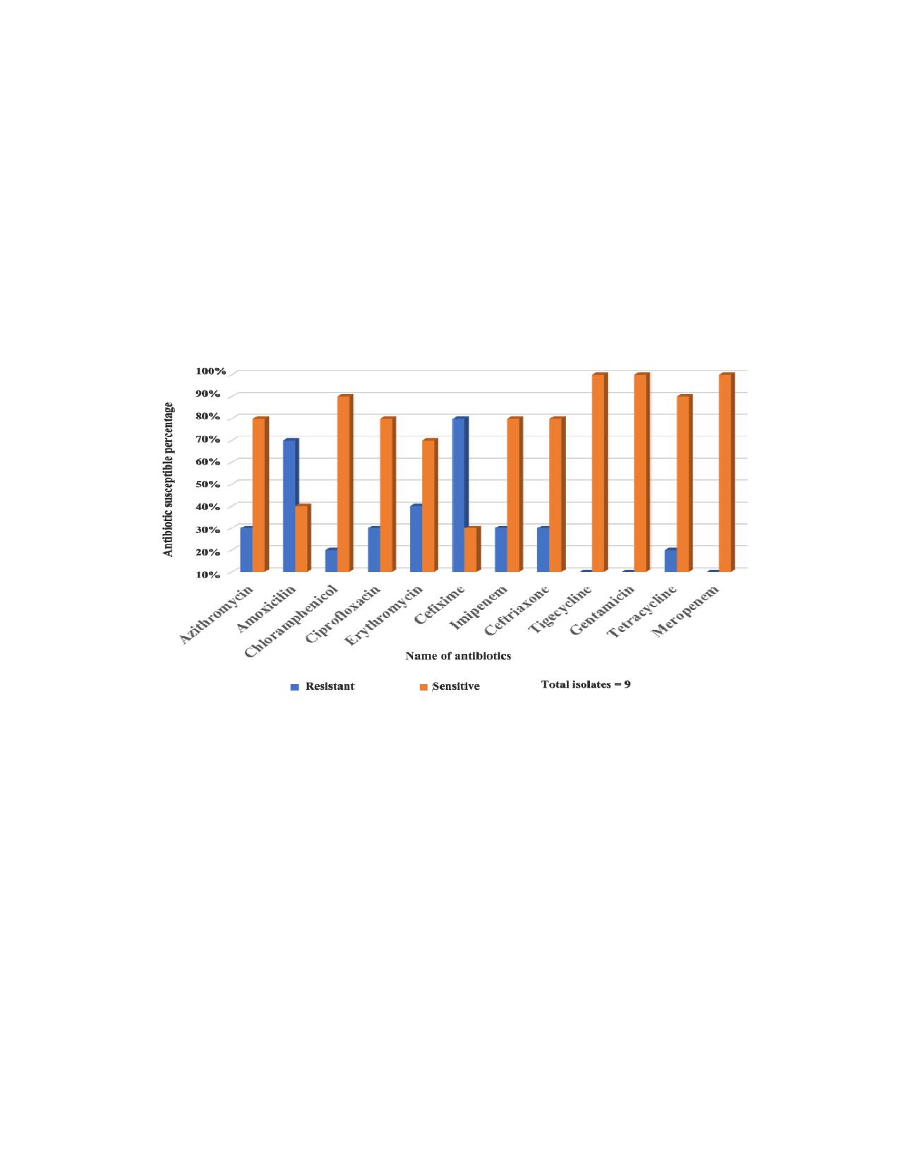
Antibiotic Susceptibility Analysis of Klebsiella Pneumoniae:
Most of the positive Klebsiella pneumoniae isolates showed in the relatively high antibiotic
susceptibility, with the exception of Amoxicillin (68%), Cefixime (79%) which demonstrated
substantial resistance & Erythromycin (39%) comparatively lower resistance. The efficiency of
these 12 antibiotics against the isolates is displayed in Figure 4(b) below. From the graph, it is
evident that Ciprofloxacin, Azithromycin, Gentamicin, Tetracycline, Tigecycline and Meropenem
showed high susceptibility, with 75% to 98% of the isolates being susceptible.
Figure 4(b): Antibiotic Susceptibility Klebsiella pneumoniae. The total number of isolates was 9
Antibiotic Susceptibility Analysis of Escherichia coli:
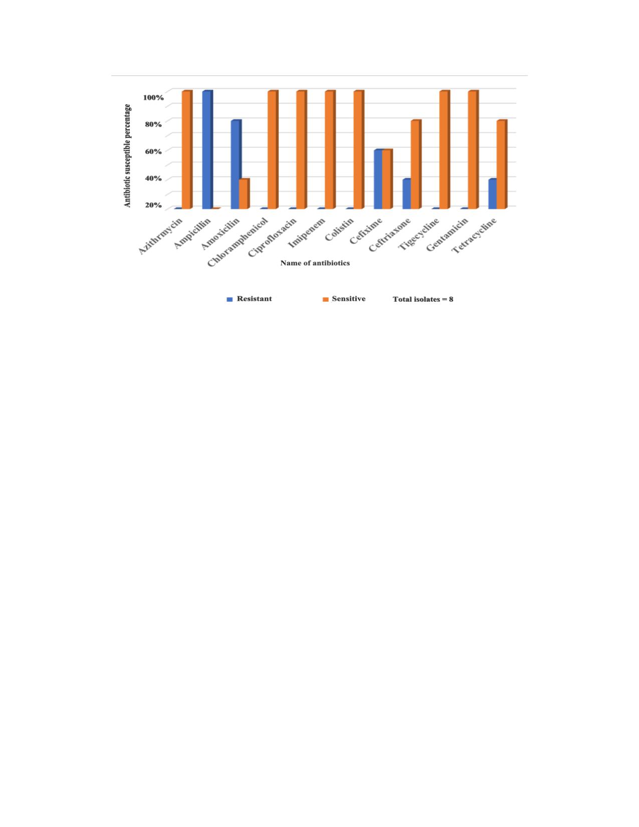
Out of the all-positive Escherichia coli isolates showed relatively high antibiotic susceptibility,
with the exception of Amoxicillin (80%) and Ampicillin (99%), which demonstrated substantial
resistance. Cefixime (58%), Ceftriaxone (28%) and Tetracycline (38%) resistance of positive
isolates. The efficiency of these 12 antibiotics against the isolates is displayed in the graph Figure
4(c) below. From the graph it is evident that Azithromycin, Chloramphenicol, Ciprofloxacin,
Imipenem, Colistin, Tigecycline, Gentamicin showed high susceptibility, with 100% of the isolates
being susceptible.
31
Figure 4(c): Antibiotic Susceptibility Profiling of Escherichia coli. The total number of isolates
was 8.
Antibiotic Susceptibility Analysis of Staphylococcus aureus:
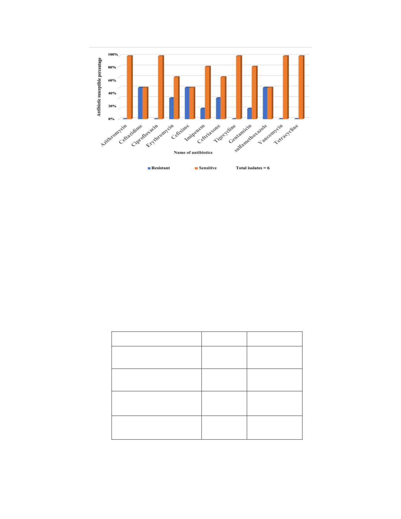
Out of all positive Staphylococcus aureus isolates showed relatively high antibiotic susceptibility,
with the exception of Cefixime, Sulfamethoxazole and Ceftazidime showed 49% of resistance.
Then Erythromycin and Ceftriaxone showed 28% resistance among positive isolates. Imipenem
and Gentamicin showed 19% of resistance which is comparatively lower. The efficiency of these
12 antibiotics against the isolates is displayed in the graph Figure 4(d) below. From the graph it
is evident that Azithromycin, Ciprofloxacin, Tigecycline, Vancomycin, Tetracycline and
Imipenem showed high susceptibility, with 85% to 100% of the Positive isolates
32
Figure 4(d): The Graph showing the resistance and sensitivity to various antibiotics of
Staphylococcus aureus. The total number of isolates was 6
3.4: Multi-drug resistant bacteria
Among the all-positive isolates of Salmonella typhi shows 16.67% for MDR & 2.32% for XDR.
MDR in klebsiella pneumoniae isolates showed 66.67% with 11% XDR. Escherichia coli has
shown MDR in 25% but no XDR. Lastly, gram positive bacteria Staphylococcus aureus showed
MDR in 50% & 16.67% XDR. The graph (Figure 5) shown MDR and XDR pattern below,
Organism
MDR
XDR
Salmonella typhi(N=43)
7 (16.67%)
1(2.32%)
Escherichia coli(N=8)
2 (25%)
0
Klebsiella
6 (66.67%)
1(11%)
pneumoniae(N=9)
Staphylococcus
3 (50%)
1 (16.67%)
aureus(N=6)
Figure 5: The table showing MDR and XDR pattern
33
CHAPTER 4
Discussion
This study aimed to assess the bacteriological profile and antibiotic resistance patterns of
septicemia pathogens among diagnosed patients at the four branches of Islamic Bank Hospital in
Dhaka city, Bangladesh. The study's findings revealed that among a total of 80 samples,
Salmonella typhi was the most prevalent pathogen, accounting for 65.2% of the isolates, followed
by Klebsiella pneumoniae (13.6%), Escherichia coli (12.1%), and Staphylococcus aureus (9.1%).
This study aligns with the international and local patterns of Salmonella typhi, Klebsiella
pneumoniae, Escherichia coli, and Staphylococcus aureus being the predominant pathogens in
bloodstream infections (BSIs). The prevalence of Salmonella typhi in this study (65.2%) is
consistent with findings from Kathmandu, Nepal, where Salmonella typhi accounted for 71% of
blood culture isolates (Simkhada, 2016). The high prevalence of Salmonella typhi in both studies
could be attributed to the common occurrence of typhoid fever in countries with poor sanitation
and water contamination issues. Typhoid fever, which has not yet been eradicated, continues to
cause septicemia (Simkhada, 2016); therefore, it is not surprising that in this study Salmonella
typhi was the leading pathogen with 65.2% of the isolates. Dagnew et al. (2013) reported similar
results in Ethiopia and Nepal, where Salmonella typhi dominated the septicemia pathogen.
The prevalence of klebsiella pneumoniae in our study (13.6%) is much lower compared to the
reported for Sub-Saharan Africa (54.4%). Escherichia Coli in your study (12.1%) is slightly lower
than the study reported in Sub-Saharan Africa (18.4%) (Lester et al., 2019). According to our
analysis, the prevalence of Staphylococcus aureus was strikingly low at 9.1%, unlike other studies
conducted in Bangladesh which noted a higher prevalence where Staphylococcus aureus was more
often associated with septicemia. More specifically, this is one of the comparisons that can be
made regarding the use of bacterial infections in the current study and Staphylococcus aureus for
past investigations conducted in the same part of the world. In contrast to the studies conducted
in Mymensingh, Bangladesh, which reported a prevalence of approximately 20%, the current study
showed a lower prevalence of 9.1%. This is likely due to differences in hospital-based settings or
the specific antibiotic resistance trends governing the prevalence of Staphylococcus aureus
34
infections in the community. Various reasons explain this, including differences in the patient
populations, healthcare settings, or methodologies employed in the studies (Khan et al., 2024).
The study analysis showed that septicemia was primarily in the elderly (60 years and above) which
contributed to 43.75% of the cases followed by the 40-59 age group with 25%. This aligns with
other global reviewed article where in older adults (≥65) constitute 58-65% of sepsis cases (Starr
& Saito, 2014). Due to septicemia being more common amongst older adults because of the
immunosenescence- a weakening immune system due to aging-associated with heightened
vulnerability to infections (Opal et al., 2005). Therefore, the geriatric population continues to be
at risk to certain community infections notably caused by Salmonella typhi, Escherichia coli, and
Klebsiella pneumoniae which could lead to potentially devastating complications if they are not
treated promptly. On the basis of gender, the males in this study outnumbered the females, 56.25%
and 43.75% respectively. Male predominance in septicemia cases is consistently reported globally,
In Wuhan, study found 61% male, 39% female among 2,111 sepsis patients (Zhao et al., 2025).
This disparity also aligns with other studies which demonstrate that men are at higher risk of
septicemia because of the differences in immune responses (Pradipta et al., 2013). It is known that
women’s immune response is enhanced due to the presence of estrogens, thus giving women better
defenses against infections, particularly in community settings.
This study highlights the increasing trend of antimicrobial resistance in Bangladesh. Antimicrobial
resistance (AMR) remains a critical global health challenge, with developing countries like
Bangladesh facing particularly high rates due to factors such as unregulated antibiotic use, and
inadequate healthcare infrastructure. In this study, Salmonella typhi (65.2%), Klebsiella
pneumoniae (13.6%), and Escherichia coli (12.1%) were the most prevalent pathogens in
septicemia patients, reflecting similar patterns observed in other countries. For instance, in Nepal,
Salmonella typhi was responsible for 71% of bloodstream infections (Simkhada, 2016), while in
Ethiopia, Klebsiella pneumoniae and Escherichia coli were also leading causes of sepsis, with
resistance rates of 68% to amoxicillin and 79% to cefixime (Dagnew et al., 2013). In our studies,
Salmonella typhi showed resistance to nalidixic acid (89%) and ampicillin (79%). Resistance to
commonly used antibiotics like ampicillin, azithromycin, and nalidixic acid in this study mirrors
trends in India, where Klebsiella pneumoniae and Escherichia coli show similar resistance patterns
to beta-lactams and fluoroquinolones (Khan et al., 2022). However, the study found relatively low
35
prevalence of Staphylococcus aureus (9.1%), which contrasts with other studies in Bangladesh
and globally, where Staphylococcus aureus typically accounts for a larger portion of septicemia
cases (Khan et al., 2024). Additionally, colistin resistance observed in the study aligns with rising
concerns in countries like China and India, where colistin resistance is becoming an emerging
public health threat, especially in multidrug-resistant Gram-negative infections (Zhu et al., 2021).
In contrast, in this study colistin is a more effective antibiotic against Salmonella typhi, Klebsiella
pneumoniae, Escherichia coli, and Staphylococcus aureus. While resistance rates in high-income
countries like the U.S. are comparatively lower, the increasing global spread of MDR and XDR
pathogens highlights the urgency of implementing robust infection control, better surveillance
systems, and effective antibiotic stewardship programs worldwide (Smith et al., 2023). Multidrug-
resistant (MDR) pathogens are a growing global concern, as demonstrated by the high prevalence
of MDR Salmonella typhi, Klebsiella pneumoniae, Escherichia coli, and Staphylococcus aureus
in this study. Isolates of 16.67% Salmonella typhi were identified as MDR, with 2.32% classified
as extensive drug-resistant (XDR). Additionally, Klebsiella pneumoniae isolates were shown
66.67% of MDR, with 11% were XDR, while Escherichia coli isolates were shown 25% of MDR,
and Staphylococcus aureus isolates were shown 50% of MDR, with 16.67% of XDR. Similar
findings have been reported in other countries, such as India, where 50% of Salmonella typhi
isolates were MDR (Khan et al., 2022), and Ethiopia, where 40% of Klebsiella pneumoniae
isolates were resistant to multiple drugs (Dagnew et al., 2013). In contrast, the prevalence of MDR
strains in developed countries like the U.S. is relatively lower, with Klebsiella pneumoniae MDR
rates reported around 15-20% (Smith et al., 2023). MDR rate reported in a study from south India,
where 24.6% of Escherichia coli isolates from bloodstream infections showed multidrug resistance
(Babu et al., 2018). The global rise in MDR and extensive drug-resistant (XDR) pathogens
highlights the need for improved infection control, antibiotic stewardship, and timely diagnostics
to manage AMR effectively in both high-income and low-income countries (Bayot & Bragg,
2024).
36
CHAPTER 5
Conclusion
Our studies have shown that most of the pathogens are already resistant to antibiotics like
Amikacin, Gentamicin, Imipenem, Tigecycline, Linezolid and Vancomycin. Age, gender and
immunity might also play a role in the frequency of the onset of sepsis and the effectiveness of
antibiotics. In our study, most of the sepsis patients are more than 60 years old (43%) and
male(56.25%). The thickness of the peptidoglycan layer on the surface of bacteria may have an
impact on their pathogenicity. Our study showed that 77% of the patients were infected by gram
negative bacteria, especially Salmonella typhi. Nevertheless, data on antibiotic susceptibility is not
universal and permanent, because antibiotic susceptibility or resistance varies from strain to strain,
and the results will differ with variations in location or continent. In addition, the antibiotics that
are currently effective on a pathogen, may not retain the same efficacy after a few years. Some of
them may even gain multidrug resistance. This happens because antibiotics pose a selective
pressure on microbes, or which they have to survive by evolving with newer antibiotic resistance
genes, which spread rapidly within the microbial kingdom (through horizontal and vertical gene
transfer). Nevertheless, we need to continue a thorough longitudinal study with larger sample sizes
(annually if possible), to validate the findings of the research and get more insights.
37
CHAPTER 6
References
1.
Addgene:
Protocol
-
How
to
run
an
Agarose
gel.
(n.d.).
2.
Ahmed I, Rabbi MB, Sultana S. Antibiotic resistance in Bangladesh: A systematic review.
Int J Infect Dis. 2019 Mar;80:54-61. Antibiotic resistance in Bangladesh: A systematic
review - PubMed
3.
Ann C. Smith ,Marise A. Hussey( 2005), Gram Stain Protocols, American Society for
001de9a5ece3/gram-stain-protocol-2886.pdf
4.
Babu, M., Menon, V. P., & P, U. D. (2018). Prevalence of antimicrobial resistant
pathogens in severe sepsis and septic shock patients. Journal of Young Pharmacists,
5.
Baral, P., Afnan, N., Zahra, M. A., Akter, B., Prapti, S. R., Hossan, M. M., & Haque, F. K.
M. (2024). Bacteriological analysis and antibiotic resistance in patients with diabetic foot
ulcers
in
Dhaka.
PLoS
ONE,
19(5),
e0301767.
6.
Bayot, M. L., & Bragg, B. N. (2024, May 27). Antimicrobial susceptibility testing.
StatPearls - NCBI Bookshelf. https://www.ncbi.nlm.nih.gov/books/NBK539714/
7.
Bepari, A. K., Rabbi, G., Shaon, H. R., Khan, S. I., Zahid, Z. I., Dalal, K., & Reza, H. M.
(2023a). Factors Driving Antimicrobial Resistance in Rural Bangladesh: A Cross-Sectional
Study on Antibiotic Use-Related Knowledge, Attitude, and Practice among Unqualified
village medical Practitioners and pharmacy shopkeepers. Advances in Therapy, 40(8),
3478-3494. https://doi.org/10.1007/s12325-023-02547-5
8.
Bio-Rad Laboratories. (2011). Disks for antibiotic susceptibility testing: Codes and
interpretative
criteria.
rad.com/webroot/web/pdf/inserts/CDG/en/Literature/inserts/66098_01_2011_GB.pdf
9.
Dagnew, M., Yismaw, G., Gizachew, M., Gadisa, A., Abebe, T., Tadesse, T., Alemu, A.,
& Mathewos, B. (2013). Bacterial profile and antimicrobial susceptibility pattern in
38
septicemia suspected patients attending Gondar University Hospital, Northwest Ethiopia.
10. Dashtian, K., Gheshlaghi, F. A., Zare-Dorabei, R., & Mahdavi, M. (2024). Prussian Blue
Analogues-Derived Molecularly Imprinted nanozyme array for septicemia detection. ACS
11. Garbern, S. C., Mamun, G. M. S., Shaima, S. N., Hakim, N., Wegerich, S., Alla, S., Sarmin,
M., Afroze, F., Sekaric, J., Genisca, A., Kadakia, N., Shaw, K., Rahman, A. S. M. M. H.,
Gainey, M., Ahmed, T., Chisti, M. J., & Levine, A. C. (2024). A novel digital health
approach to improving global pediatric sepsis care in Bangladesh using wearable
technology and machine learning. PLOS Digital Health,
3(10), e0000634.
12. Gogoi, M., Chetia, P., Pegu, R. K., & Roy, J. D. (2024). Targeting nuc Gene for Detection
of Staphylococcus aureus from Bovine Milk Samples of Assam, India and Phenotypic
Identification of Antibiotic Resistance - A New Insight. Journal of Pure and Applied
13. Gücer, L. S., Pınarlık, F., Farzana, R., Ataç, N., Genç, Z., Madran, B., Bulbul, I. S., Şahin,
F., Sengel, B. E., Tigen, E. T., Ilki, A., Sari, N., Azap, Ö. K., Can, F., Walsh, T. R., &
Ergönül, Ö. (2024). Antimicrobial resistance rates and treatment options in bloodstream
infections: a prospective observational study. Journal of Global Antimicrobial Resistance,
14. Green, M. R., & Sambrook, J. (2012). Molecular Cloning: A Laboratory Manual (4th ed.).
Cold Spring Harbor Laboratory Press.
15. Halim, M. R., & Nishat, R. J. (2024). Opportunities and challenges of application of
cytosorb therapy in sepsis management. Bangladesh Critical Care Journal, 12(2), 79-80.
16. Jan Hudzicki. (2009). Kirby-Bauer Disk Diffusion Susceptibility Test Protocol. In
AMERICAN SOCIETY FOR MICROBIOLOGY. Retrieved May
15,
2025, from
disk-diffusion-susceptibility-test-protocol-pdf.pdf
17. Khan MS, Mamun AA, Khan MSI, Sathi FA, Paul SK, Nasrim SA, Haque N, Arafa P, Nila
SS, Roy S, Nahar A. Antimicrobial Susceptibility Patterns of Bacterial Isolates from Blood
39
Culture of Pediatric Patients with Suspected Sepsis at a Tertiary Care Hospital in
Mymensingh, Bangladesh. Mymensingh Med J. 2024 Jan;33(1):49-61. Antimicrobial
Susceptibility Patterns of Bacterial Isolates from Blood Culture of Pediatric Patients with
Suspected Sepsis at a Tertiary Care Hospital in Mymensingh, Bangladesh - PubMed
18. Kingsley, O. C., Ifeanyi, A. O., Edet, A. E., & Smart, O. C. (2013). Bacteriologic profile
and antibiotic susceptibility pattern of suspected septicaemic patients in Uyo, Nigeria. Res
J Med Sci, 7(2), 35-39.
19. Leung, L. Y., Huang, H., Hung, K. K., Leung, C. Y., Lam, C. C., Lo, R. S., Yeung, C. Y.,
Tsoi, P. J., Lai, M., Brabrand, M., Walline, J. H., & Graham, C. A. (2024). Door-to-
antibiotic time and mortality in patients with sepsis: Systematic review and meta-analysis.
European
Journal
of
Internal
Medicine,
129,
48-61.
20. Lester, R., Musicha, P., Van Ginneken, N., Dramowski, A., Hamer, D. H., Garner, P., &
Feasey, N. A. (2019). Prevalence and outcome of bloodstream infections due to third-
generation cephalosporin-resistant Enterobacteriaceae in sub-Saharan Africa: a systematic
review.
Journal
of
Antimicrobial
Chemotherapy,
75(3),
492-507.
21. Lindsey, R. L., Garcia-Toledo, L., Fasulo, D., Gladney, L., & Strockbine, N. (2017).
Multiplex polymerase chain reaction for identification of Escherichia coli , Escherichia
albertii and Escherichia fergusonii. Journal of Microbiological Methods,
140,
1-4.
22. Lorenz, T. C. (2012). Polymerase chain Reaction: basic protocol plus troubleshooting and
optimization
strategies.
Journal
of
Visualized
Experiments,
63.
23. Moore, Edward & Arnscheidt, A. & Krüger, A. & Strömpl, Carsten & Mau, M.. (2004).
Simplified protocols for the preparation of genomic DNA from bacterial cultures.
24. Opal, S. M., Girard, T. D., & Ely, E. W. (2005). The immunopathogenesis of sepsis in
elderly patients. Clinical Infectious Diseases,
41(Supplement
7), S504-S512.
25. Pradipta, I., Sodik, D., Parwati, I., Lestari, K., Halimah, E., Diantini, A., & Abdulah, R.
(2013). Antibiotic resistance in sepsis patients: Evaluation and recommendation of
40
antibiotic use. North American Journal of Medical Sciences,
5(6),
344.
26. Prakash Simkhada & Tribhuvan University, Nepal. (2016). Bacteriological Profile and
Antibiotic Susceptibility Pattern of Blood Culture Isolates from Patients Visiting Tertiary
Care Hospital in Kathmandu, Nepal. Global Journal of Medical Research: C Microbiology
and
Pathology,
16(1).
Shrestha/publication/305205543_Bacteriological_Profile_and_Antibiotic_Susceptibility_
Pattern_of_Blood_Culture_Isolates_from_Patients_Visiting_Tertiary_Care_Hospital_in_
Kathmandu_Nepal/links/57849a0a08ae37d3af6c24d9/Bacteriological-Profile-and-
Antibiotic-Susceptibility-Pattern-of-Blood-Culture-Isolates-from-Patients-Visiting-
Tertiary-Care-Hospital-in-Kathmandu-Nepal.pdf
27. Ranjbar, R., Naghoni, A., Afshar, D., Nikkhahi, F., & Mohammadi, M. (2016). Rapid
Molecular Approach for Simultaneous Detection of Salmonella spp., Shigella spp., and
Vibrio cholera. Osong Public Health and Research Perspectives, 7(6), 373-377.
28. Saiki, R. K., et al. (1988). Primer-directed enzymatic amplification of DNA with a
thermostable
DNA
polymerase.
Science,
239(4839),
487-491.
29. Takara Bio Inc. EmeraldAmp PCR Master Mix Product Manual.
31. Washington JA 2nd, Yu PK, Gavan TL, Schoenknecht FD, Thornsberry C. Interpretation
of the disk diffusion susceptibility test for amikacin: report of a collaborative study.
Antimicrob
Agents
Chemother.
1979
Mar;15(3):400-
32. Zahra, M. A., Toma, T. T., Nasreen, S., Zarin, Z. T. R., Khan, Z. T. S. E., & Haque, F. K.
M.
(2025). Characterizing the co-existence of metallo-β-lactamase-producing and
extended spectrum β-lactamase-producing Escherichia coli and Klebsiella pneumoniae
isolates in community wastewater samples of Dhaka, Bangladesh. Journal of Water and
41
33. Zhao, H., Yang, B., Dai, H., Li, C., Ruan, H., & Li, Y. (2025). Sex Differences in Sepsis-
Related Acute Respiratory Distress Syndrome and Other Short-Term Outcomes among
Critically Ill Patients with Sepsis: A Retrospective Study in China. Shock.
34. Zhu, X.-Q., Liu, Y.-Y., Wu, R., Xun, H., Sun, J., Li, J., Feng, Y., & Liu, J.-H. (2021).
Impact of mcr-1 on the development of high-level colistin resistance in Klebsiella
pneumoniae and Escherichia coli. Frontiers in Microbiology,
12,
666782.