AHA/ACC SCIENTIFIC STATEMENT (2024) - page 9
March 2024
AGA Clinical Practice Update on Diet and IBD
529
feel that nutrition is either not being prioritized in their care
and intestinal remodeling resulting from gut inflammation,
or is being overlooked, and often report that dietary advice
patients with IBD can be instructed to modify the texture of
from non-RD practitioners is heterogenous and conflict-
fibrous foods by cooking, blending, and thoroughly chewing
ing.69
This results in self-directed nutrition changes,
fruits and vegetables, which will allow them to better
resulting in unnecessary dietary restriction and increased
tolerate a healthy diet over the lifespan.
risk of developing malnutrition.57 RDs are fundamental
When there is active inflammation and/or stricturing com-
members of the multidisciplinary IBD care team, as they
plications of CD, liquid nutrition formulas have demonstrated
have expertise in diagnosis and management of malnutri-
efficacy. Complete dietary modification with EEN using
tion, including specific protein, macronutrient, vitamin, and
commercially available liquid nutrition formulas may induce
micronutrient requirements, which vary in different phases
remission in CD, with the strongest evidence in children. The
of illness.70 Ideally, referral to an RD should not be deferred
complete avoidance of regular food for a prolonged time period
until the consequences of uncontrolled disease, such as
is challenging for many patients with IBD, but clinical remission
malnutrition, become unmanageable. RDs can also play a
can be maintained with a staged reintroduction of foods. In
key role in prevention of extraintestinal complications of
patients with CD and obstructive complications necessitating
IBD, including dietary modification to prevent enteric
surgery, preoperative therapy with liquid nutrition can improve
hyperoxaluria and development of kidney stones.
nutritional status and improve operative outcomes.
PN and EN support requires a specialized practice that
Intravenous nutrition with fluids, macronutrients, vita-
necessitates the expertise of an RD. The role of the RD is to
mins, and minerals maintains an important, lifesaving role
assist the multidisciplinary team with appropriate nutrition
in the short-term management of patients with IBD during
support route selection; prescribing adequate energy, pro-
acute inflammatory and obstructive complications when the
tein, and micronutrient needs; monitoring patients
gut is no longer functioning and able to safely handle oral
throughout the duration of the treatment; adjusting the
intake. Patients with IBD with SBS will benefit from home
nutrition prescription when indicated; educating the patient
fluid and nutritional support and treatment with glucagon-
on safe administration and compliance with EN and PN
like peptide-2 medications, which enhance intestinal adap-
support, as well as guiding the patients and their care team
tation, and weaning from PN should be considered in long-
on the appropriate transition back to a regular oral intake.
term management.
Best Practice Advice 12: Breastfeeding is associ-
Dietary advice needs to be tailored to an individual IBD
ated with a lower risk for diagnosis of IBD during
patient’s nutritional status and goals, which will vary over
childhood. A healthy, balanced, Mediterranean diet rich
time. Implementing the more complex nutritional strategies
in a variety of fruits and vegetables and decreased
for IBD management will be best achieved by means of
intake of ultraprocessed foods have been associated
collaborative interdisciplinary practice between gastroen-
with a lower risk of developing IBD.
terologists and RDs.
Numerous studies have investigated potential predis-
posing and protective factors for developing IBD. Such
References
studies focus on early life exposures, including maternal
1. Lewis JD, Sandler RS, Brotherton C, et al. A randomized
health, in utero exposures, mode of delivery, breastfeeding,
trial comparing the specific carbohydrate diet to a
different diets, and antibiotic exposure, among many other
Mediterranean diet in adults with Crohn’s disease.
factors. Breastfeeding has been associated with a decreased
Gastroenterology 2021;161:837-852.e9.
risk of developing both CD and UC and the protective effect
2. Chicco F, Magri S, Cingolani A, et al. Multidimensional
was strongest for the group who breastfed for at least 12
impact of Mediterranean diet on IBD patients. Inflamm
months.71,72 Regarding diet, results have been conflicting
Bowel Dis 2021;27:1-9.
and controversial. The healthy, balanced, Mediterranean
3. Cox SR, Linday JO, Fromentin S, et al. Effects of low
diet is the preferred diet for almost all individuals. Con-
FODMAP diet on symptoms, fecal microbiome, and
sumption of high levels of ultraprocessed foods and low
markers of inflammation in patients with quiescent in-
intake of dietary fiber from fruits and vegetables have been
flammatory bowel disease in a randomized trial.
associated with an increased risk of developing CD.68,73,74
Gastroenterology 2020;158:176-188.e7.
4. Ng SC, Benjamin JL, McCarthy NE, et al. Relationship
Conclusions
between human intestinal dendritic cells, gut microbiota,
There have been significant advances in our under-
and disease activity in Crohn’s disease. Inflamm Bowel
standing of the importance of diet and nutrition in IBD, not
Dis 2011;17:2027-2037.
only in disease etiopathogenesis, but more recently as a
5. Sokol H, Pigneur B, Watterlot L, et al. Faecalibacterium
disease-modifying factor and treatment. A healthy, balanced
prausnitzii is an anti-inflammatory commensal bacterium
Mediterranean-style diet rich in fruits and vegetables,
identified by gut microbiota analysis of Crohn disease
complex carbohydrates, and monounsaturated fats and low
patients. Proc Natl Acad Sci U S A
2008;
in added sugar, salt, red meats, and ultraprocessed foods
105:16731-16736.
can be tolerated by many patients and may improve
6. Varela E, Manichanh C, Gallart M, et al. Colonisation by
symptoms in some. A Mediterranean-style diet can be
Faecalibacterium prausnitzii and maintenance of clinical
introduced when patients are asymptomatic or when having
remission in patients with ulcerative colitis. Aliment
mild to moderate symptoms. Due to the stricturing damage
Pharmacol Ther 2013;38:151-161.
530
Hashash et al
Gastroenterology Vol. 166, Iss. 3
7.
Chen J, Wellens J, Kalla R, et al. Intake of ultra-
Crohn’s disease: clinical and nutritional outcome. Gut
processed foods is associated with an increased risk of
1994;35:783-787.
Crohn’s disease: a cross-sectional and prospective
24.
Takagi S, Utsunomiya K, Kuriyama S, et al. Effectiveness
analysis of 187 154 participants in the UK Biobank.
of an ’half elemental diet’ as maintenance therapy for
J Crohns Colitis 2023;17:535-552.
Crohn’s disease: a randomized-controlled trial. Aliment
8.
Lo CH, Khandpur N, Rossato SL, et al. Ultra-processed
Pharmacol Ther 2006;24:1333-1340.
foods and risk of Crohn’s disease and ulcerative colitis: a
25.
Johnson T, Macdonald S, Hill SM, et al. Treatment of
prospective cohort study. Clin Gastroenterol Hepatol
active Crohn’s disease in children using partial enteral
2022;20:e1323-e1337.
nutrition with liquid formula: a randomised controlled
9.
Fu T, Chen H, Chen X, et al. Sugar-sweetened beverages,
trial. Gut 2006;55:356-361.
artificially sweetened beverages and natural juices and risk
26.
Yanai H, Levine A, Hirsch A, et al. The Crohn’s disease
of inflammatory bowel disease: a cohort study of 121,490
exclusion diet for induction and maintenance of remis-
participants. Aliment Pharmacol Ther 2022;56:1018-1029.
sion in adults with mild-to-moderate Crohn’s disease
10.
Ahsan M, Koutroumpakis F, Ramos Rivers C, et al. High
(CDED-AD): an open-label, pilot, randomised trial. Lancet
sugar-sweetened beverage consumption is associated
Gastroenterol Hepatol 2022;7:49-59.
with increased health care utilization in patients with in-
27.
Niseteo T, Sila S, Trivic I, et al. Modified Crohn’s disease
flammatory bowel disease: a multiyear, prospective
exclusion diet is equally effective as exclusive enteral
analysis. J Acad Nutr Diet 2022;122:1488-1498.e1.
nutrition: real-world data. Nutr Clin Pract
2022;
11.
Crohn’s and Colitis Canada. Diet and nutrition. Available at:
37:435-441.
28.
Herrador-López M, Martín-Masot R, Navas-López VM.
Journey/Diet-and-Nutrition-in-IBD. Accessed September
EEN yesterday and today . . . CDED today and tomorrow.
2, 2023.
Nutrients 2020;12(12):3793.
12.
Crohn’s and Colitis Foundation. Diet and nutrition.
29.
Levine A, Wine E, Assa A, et al. Crohn’s disease exclu-
Available at: https://www.crohnscolitisfoundation.org/
sion diet plus partial enteral nutrition induces sustained
diet-and-nutrition. Accessed September 2, 2023.
remission in a randomized controlled trial. Gastroenter-
13.
Serrano Fernandez V, Palomino MS, Laredo-Aguilera JA,
ology 2019;157:440-450.e8.
et al. High-fiber diet and Crohn’s disease: systematic
30.
Sigall Boneh R, Sarbagili Shabat C, Yanai H, et al. Dietary
review and meta-analysis. Nutrients 2023;15(14):3114.
therapy with the Crohn’s disease exclusion diet is a
14.
Heaton KW, Thornton JR, Emmett PM. Treatment of
successful strategy for induction of remission in children
Crohn’s disease with an unrefined-carbohydrate, fibre-
and adults failing biological therapy. J Crohns Colitis
rich diet. Br Med J 1979;2(6193):764-766.
2017;11:1205-1212.
15.
Lamb CA, Kennedy NA, Raine T, et al. British Society of
31.
Triantafillidis JK, Vagianos C, Papalois AE. The role of
Gastroenterology consensus guidelines on the man-
enteral nutrition in patients with inflammatory bowel
agement of inflammatory bowel disease in adults. Gut
disease: current aspects. Biomed Res Int 2015;2015:
2019;68(Suppl 3):s1-s106.
197167.
16.
Forbes A, Escher J, Hébuterne X, et al. ESPEN guideline:
32.
Meade S, Patel KV, Luber RP, et al. A retrospective
clinical nutrition in inflammatory bowel disease. Clin Nutr
cohort study: pre-operative oral enteral nutritional opti-
2017;36:321-347.
misation for Crohn’s disease in a UK tertiary IBD centre.
17.
Whitten KE, Rogers P, Ooi CKY, et al. International sur-
Aliment Pharmacol Ther 2022;56:646-663.
vey of enteral nutrition protocols used in children with
33.
Impact of malnutrition on early outcomes after cancer
Crohn’s disease. J Dig Dis 2012;13:107-112.
surgery: an international, multicentre, prospective cohort
18.
Heuschkel RB. Enteral nutrition in children with Crohn’s
study. Lancet Glob Health 2023;11:e341-e349.
disease. J Pediatr Gastroenterol Nutr 2000;31:575.
34.
Costa-Santos MP, Palmela C, Torres J, et al. Preopera-
19.
Melton SL, Fitzpatrick JA, Taylor KM, et al. Lessons from
tive enteral nutrition in adults with complicated Crohn’s
an audit of exclusive enteral nutrition in adult inpatients
disease: effect on disease outcomes and gut microbiota.
and outpatients with active Crohn’s disease: a single-
Nutrition 2020;70s:100009.
centre experience. Frontline Gastroenterol 2023;14:6-12.
35.
Rocha A, Bessa I, Lago P, et al. Preoperative enteral
20.
Rigaud D, Cosnes J, Le Quintrec Y, et al. Controlled trial
nutrition and surgical outcomes in adults with Crohn’s
comparing two types of enteral nutrition in treatment of
disease: a systematic review. GE Port J Gastroenterol
active Crohn’s disease: elemental versus polymeric diet.
2019;26:184-195.
Gut 1991;32:1492-1497.
36.
Detsky AS, Baker JP, O’Rourke K, et al. Predicting
21.
Kakkadasam Ramaswamy P. Exclusive enteral nutrition
nutrition-associated complications for patients under-
with oral polymeric diet helps in inducing clinical and
going gastrointestinal surgery. JPEN J Parenter Enteral
biochemical remission in adults with active Crohn’s
Nutr 1987;11:440-446.
disease. JPEN J Parenter Enteral Nutr 2022;46:423-432.
37.
Heyland DK, Montalvo M, MacDonald S, et al. Total
22.
Wall CL, Gearry RB, Day AS. Treatment of active Crohn’s
parenteral nutrition in the surgical patient: a meta-anal-
disease with exclusive and partial enteral nutrition: a pilot
ysis. Can J Surg 2001;44:102-111.
study in adults. Inflamm Intest Dis 2018;2:219-227.
38.
Nelson AD, Elkins JR, Stocchi L, et al. Use and misuse of
23.
Royall D, Jeejeebhoy KN, Baker JP, et al. Comparison of
parenteral nutrition in patients with inflammatory bowel
amino acid v peptide based enteral diets in active
disease. Inflamm Bowel Dis 2022;28:1592-1602.
March 2024
AGA Clinical Practice Update on Diet and IBD
531
39.
Braunschweig CL, Levy P, Sheean PM, et al. Enteral
56.
Bischoff SC, Escher J, Hébuterne X, et al. ESPEN
compared with parenteral nutrition: a meta-analysis. Am
practical guideline: clinical nutrition in inflammatory
J Clin Nutr 2001;74:534-542.
bowel disease. Clin Nutr 2020;39:632-653.
40.
Mueller C. The ASPEN Adult Nutrition Support Core
57.
Casanova MJ, Chaparo M, Molina B, et al. Prevalence of
Curriculum. 3rd ed. American Society for Parenteral and
malnutrition and nutritional characteristics of patients
Enteral Nutrition; 2017.
with inflammatory bowel disease. J Crohns Colitis 2017;
41.
Worthington P, Balint J, Bechtold M, et al. When is
11:1430-1439.
parenteral nutrition appropriate? JPEN J Parenter Enteral
58.
Cederholm T, Jensen GL, Correia MITD, et al. GLIM criteria
Nutr 2017;41:324-377.
for the diagnosis of malnutrition - a consensus report from
42.
Guglielmi FW, Boggio-Bertinet D, Federico A, et al. Total
the global clinical nutrition community. Clin Nutr 2019;38:1-9.
parenteral nutrition-related gastroenterological compli-
59.
Taylor LM, Eslamparast T, Farhat K, et al. Using patient
cations. Dig Liver Dis 2006;38:623-642.
completed screening tools to predict risk of malnutrition
43.
Luman W, Shaffer JL. Prevalence, outcome and asso-
in patients with inflammatory bowel disease. Crohns
ciated factors of deranged liver function tests in patients
Colitis 360 2021;3(3):otab043.
on home parenteral nutrition. Clin Nutr 2002;21:337-343.
60.
Gold SL, Manning L, Kohler D, et al. Micronutrients and
44.
Iyer K, DiBaise JK, Rubio-Tapia A. AGA clinical practice
their role in inflammatory bowel disease: function,
update on management of short bowel syndrome:
assessment, supplementation, and impact on clinical
expert review. Clin Gastroenterol Hepatol
2022;
outcomes including muscle health. Inflamm Bowel Dis
20:2185-2194.e2.
2023;29:487-501.
45.
Massironi S, Cavalcoli F, Rausa E, et al. Understanding
61.
MacMaster MJ, Damianopoulou S, Thomson C, et al.
short bowel syndrome: current status and future per-
A prospective analysis of micronutrient status in quiescent
spectives. Dig Liver Dis 2020;52:253-261.
inflammatory bowel disease. Clin Nutr 2021;40:327-331.
46.
Gajendran M, Umapathy C, Loganathan P, et al. Analysis of
62.
Abraham BP, Prasad P, Malaty HM. Vitamin D deficiency
hospital-based emergency department visits for inflamma-
and corticosteroid use are risk factors for low bone
tory bowel disease in the USA. Dig Dis Sci 2016;61:389-399.
mineral density in inflammatory bowel disease patients.
47.
Pulley J, Todd A, Flatley C, et al. Malnutrition and quality
Dig Dis Sci 2014;59:1878-1884.
of life among adult inflammatory bowel disease patients.
63.
Dignass AU, Gasche C, Bettenworth D, et al. European
JGH Open 2020;4:454-460.
consensus on the diagnosis and management of iron
48.
Ananthakrishnan AN, McGinley EL. Infection-related
deficiency and anaemia in inflammatory bowel diseases.
hospitalizations are associated with increased mortality
J Crohns Colitis 2015;9:211-222.
in patients with inflammatory bowel diseases. J Crohns
64.
Evstatiev R, Marteau P, Iqbal T, et al. FERGIcor, a ran-
Colitis 2013;7:107-112.
domized controlled trial on ferric carboxymaltose for iron
49.
Ananthakrishnan AN, McGinley EL, Binion DG, et al.
deficiency anemia in inflammatory bowel disease.
A novel risk score to stratify severity of Crohn’s disease
Gastroenterology 2011;141:846-853.e1-e2.
hospitalizations. Am J Gastroenterol 2010;105:1799-1807.
65.
Hou JK, Gasche C, Drazin NZ, et al. Assessment of gaps
50.
Nguyen GC, Munsell M, Harris ML. Nationwide preva-
in care and the development of a care pathway for
lence and prognostic significance of clinically diagnos-
anemia in patients with inflammatory bowel diseases.
able
protein-calorie
malnutrition
in
hospitalized
Inflamm Bowel Dis 2017;23:35-43.
inflammatory bowel disease patients. Inflamm Bowel Dis
66.
Ward MG, Kariyawasam VC, Mogan SB, et al. Preva-
2008;14:1105-1111.
lence and risk factors for functional vitamin B12 defi-
51.
Evans DC, Corkins MR, Malone A, et al. The use of
ciency in patients with Crohn’s disease. Inflamm Bowel
visceral proteins as nutrition markers: an ASPEN position
Dis 2015;21:2839-2847.
paper. Nutr Clin Pract 2021;36:22-28.
67.
Battat R, Kopylov U, Szilagyi A, et al. Vitamin B12 defi-
52.
Yeh DD, Johnson E, Harrison T, et al. Serum levels of
ciency in inflammatory bowel disease: prevalence, risk
albumin and prealbumin do not correlate with nutrient
factors, evaluation, and management. Inflamm Bowel Dis
delivery in surgical intensive care unit patients. Nutr Clin
2014;20:1120-1128.
Pract 2018;33:419-425.
68.
Stabler SP. Clinical practice. Vitamin B12 deficiency.
53.
Marcason W. Should albumin and prealbumin be used as
N Engl J Med 2013;368:149-160.
indicators for malnutrition? J Acad Nutr Diet 2017;
69.
Holt DQ, Strauss BJ, Moore GT. Patients with inflammatory
117:1144.
bowel disease and their treating clinicians have different
54.
Fuhrman MP, Charney P, Mueller CM. Hepatic proteins
views regarding diet. J Hum Nutr Diet 2017;30:66-72.
and nutrition assessment. J Am Diet Assoc
2004;
70.
Levine A, Rhodes JM, Lindsay JO, et al. Dietary guidance
104:1258-1264.
from the International Organization for the Study of In-
55.
White JV, Guenter P, Jensen G, et al. Consensus state-
flammatory Bowel Diseases. Clin Gastroenterol Hepatol
ment: Academy of Nutrition and Dietetics and American
2020;18:1381-1392.
Society for Parenteral and Enteral Nutrition: character-
71.
Agrawal M, Sabino J, Frías-Gomes C, et al. Early life
istics recommended for the identification and docu-
exposures and the risk of inflammatory bowel disease:
mentation of adult malnutrition (undernutrition). JPEN J
systematic review and meta-analyses. EClinicalMedicine
Parenter Enteral Nutr 2012;36:275-283.
2021;36:100884.
532
Hashash et al
Gastroenterology Vol. 166, Iss. 3
72.
Xu L, Lochhead P, Ko Y, et al. Systematic review with
CRediT Authorship Contributions
Jana G. Hashash (Conceptualization: Lead; Data curation: Equal; Writing -
meta-analysis: breastfeeding and the risk of Crohn’s
original draft: Equal; Project administration: Lead; Supervision: Equal;
disease and ulcerative colitis. Aliment Pharmacol Ther
Visualization: Equal)
Jaclyn Elkins (Data curation: Equal; Writing - original draft: Equal)
2017;46:780-789.
James D. Lewis (Writing - review & editing: Equal; Supervision: Equal)
73.
Ananthakrishnan AN, Khalili H, Konijet GG, et al.
David G. Binion (Writing - original draft: Supporting; Writing - review &
A prospective study of long-term intake of dietary fiber
editing: Lead; Supervision: Equal; Visualization: Equal)
and risk of Crohn’s disease and ulcerative colitis.
Conflicts of interest
Gastroenterology 2013;145:970-977.
These authors disclose the following: James D. Lewis consulted/served on an
advisory board for Eli Lilly and Company, Samsung Bioepis, UCB, Bristol-
74.
Narula N, Chang NH, Mohammad D, et al. Food pro-
Myers Squibb, Nestlé Health Science, Merck, Celgene, Janssen
cessing and risk of inflammatory bowel disease: a sys-
Pharmaceuticals, Bridge Biotherapeutics, Entasis Therapeutics, AbbVie,
tematic review and meta-analysis. Clin Gastroenterol
Pfizer, Gilead, Arena Pharmaceuticals, Protagonist Therapeutics, Amgen,
Sanofi, and Scipher Medicine. He has had research funding from Nestlé
Hepatol 2023;21:2483-2495.e1.
Health Science, Takeda, Janssen Pharmaceuticals, and AbbVie. He has
received educational grants from Takeda and Janssen. He has performed
legal work on behalf of generic manufacturers of ranitidine, including L.
Received April 23, 2023. Accepted November 5, 2023.
Perrigo Company, Glenmark Pharmaceuticals Inc, Amneal Pharmaceuticals
LLC, Aurobindo Pharma Switzerland, Inc, Dr. Reddy’s Laboratories, Inc,
Correspondence
Novitium Pharma, Ranbaxy Inc, Sun Pharmaceutical Industries, Inc, Strides
Address correspondence to: Jana G. Hashash, MD, MSc, Division of
Pharma, Inc, and Wockhardt Switzerland LLC. He owns stock in Dark
Gastroenterology and Hepatology, Mayo Clinic,
4500
San Pablo Road,
Canyon Labs. David G. Binion has received research funding from AbbVie,
Jacksonville, Florida 32224. e-mail: AlHashash.Jana@mayo.edu.
Merck, and Takeda. The remaining authors disclose no conflicts.
Stroke
AHA SCIENTIFIC STATEMENT
Diagnosis and Management of Cerebral Venous
Thrombosis: A Scientific Statement From the
American Heart Association
Gustavo Saposnik, MD, MPH, PhD, FAHA, Chair; Cheryl Bushnell, MD, MHS, FAHA, Vice Chair; Jonathan M. Coutinho, MD, PhD;
Thalia S. Field, MD, MHSc; Karen L. Furie, MD, MPH, FAHA; Najibah Galadanci, MBBS, MPH, PhD; Wayneho Kam, MD;
Fenella C. Kirkham, MD; Norma D. McNair, RN, PhD; Aneesh B. Singhal, MD, FAHA; Vincent Thijs, MD, PhD, FAHA;
Victor X.D. Yang, MD, PhD; on behalf of the American Heart Association Stroke Council; Council on Cardiopulmonary, Critical
Care, Perioperative and Resuscitation; Council on Cardiovascular and Stroke Nursing; and Council on Hypertension
ABSTRACT: Cerebral venous thrombosis accounts for 0.5% to 3% of all strokes. The most vulnerable populations include
young individuals, women of reproductive age, and patients with a prothrombotic state. The clinical presentation of cerebral
venous thrombosis is diverse (eg, headaches, seizures), requiring a high level of clinical suspicion. Its diagnosis is based
primarily on magnetic resonance imaging/magnetic resonance venography or computed tomography/computed tomographic
venography. The clinical course of cerebral venous thrombosis may be difficult to predict. Death or dependence occurs in
10% to 15% of patients despite intensive medical treatment. This scientific statement provides an update of the 2011
American Heart Association scientific statement for the diagnosis and management of cerebral venous thrombosis. Our
focus is on advances in the diagnosis and management decisions of patients with suspected cerebral venous thrombosis.
We discuss evidence for the use of anticoagulation and endovascular therapies and considerations for craniectomy. We also
provide an algorithm to optimize the management of patients with cerebral venous thrombosis and those with progressive
neurological deterioration or thrombus propagation despite maximal medical therapy.
Key Words: AHA Scientific Statements ◼ anticoagulants ◼ headaches ◼ intracranial thrombosis ◼ seizure ◼ sinus thrombosis ◼ venous thrombosis
erebral venous thrombosis (CVT) is the presence of
This scientific statement synthetizes the clinical
a blood clot in the dural venous sinuses, the cerebral
presentation, predisposing factors, advances in imag-
C
veins, or both.1 Among those with stroke, CVT repre-
ing modalities and therapies, and management of CVT
sents only 0.5% to 3%.2 Registry-based and cohort stud-
in special populations (pediatric, pregnancy and puer-
ies suggest that CVT affects predominantly individuals
perium, and vaccine-induced CVT). This document
<55 years of age, with two-thirds occurring in women.2
is strengthened by new evidence since our previous
With regard to location, the most commonly affected
publication in 2011.1 Future areas of research are
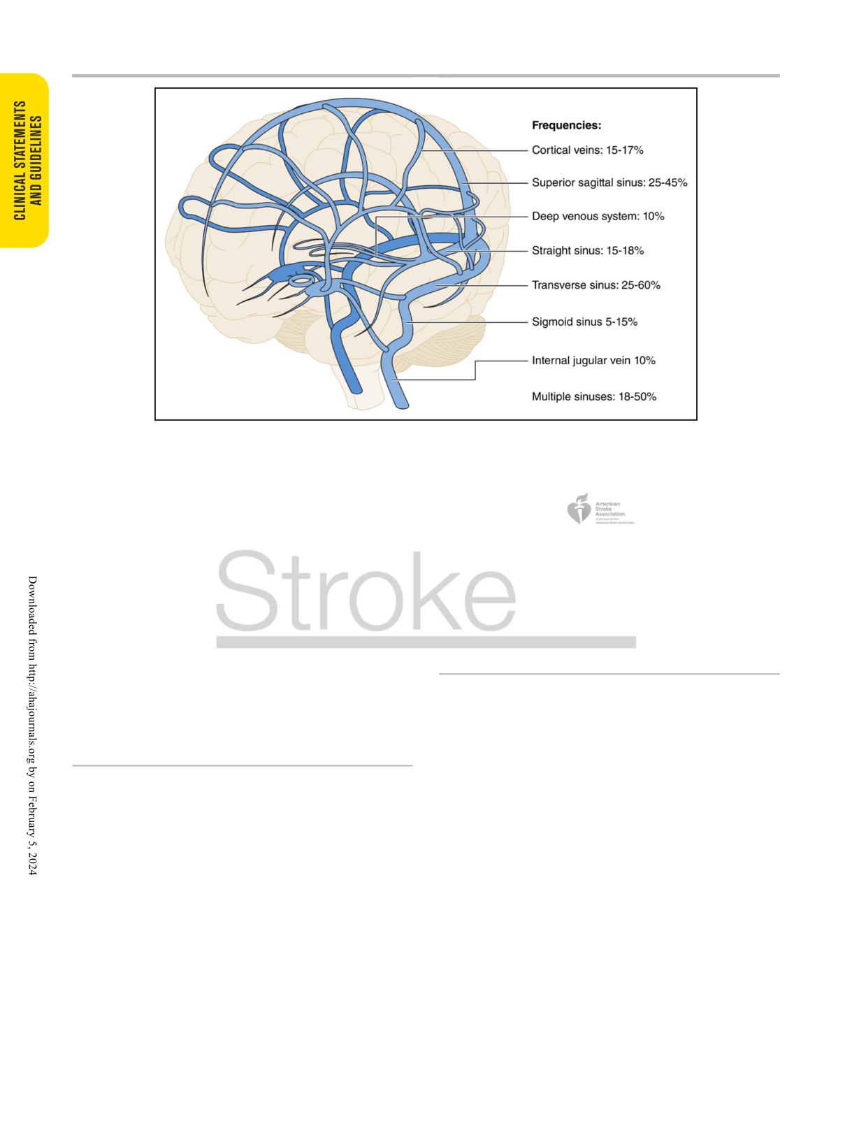
sinuses are illustrated in Figure 1.1-3
highlighted.
Overall, patients with CVT have a favorable out-
come. Most patients with CVT nowadays survive with-
CLINICAL PRESENTATION
out physical disability, but chronic symptoms, which
negatively affect quality of life, are not uncommon.
Presenting symptoms of CVT can be due to increased
Most common factors associated with poor prognosis
intracranial pressure or focal parenchymal injury, with
include advanced age, active cancer, decreased level of
or without mass effect.1 Headache is the most com-
consciousness, and intracerebral hemorrhage, among
mon symptom of CVT, occurring in nearly 90% of
others.1,4,5
cases.4 Other signs and symptoms related to intracranial
Supplemental Material is available at https://www.ahajournals.org/doi/suppl/10.1161/STR.0000000000000456.
© 2024 American Heart Association, Inc.
Stroke is available at www.ahajournals.org/journal/str
Stroke. 2024;55:e00-e00. DOI: 10.1161/STR.0000000000000456
TBD 2024 e1
Saposnik et al
Diagnosis and Management of Cerebral Venous Thrombosis
Figure 1. Anatomy of the cerebral venous system and distribution of CVT.
Prevalence of sinus involvement in CVT. Percentages may be >100% because many patients may have >1 sinus involved.1-3 Please note that
internal jugular vein thrombosis represents its concomitant prevalence with CVT (not in isolation). CVT indicates cerebral venous thrombosis.
pressure include nausea, transient visual obscurations
can be associated with CVT.1,4,7,16 Other transient provoking
or vision loss (13%-27%), papilledema, and diplopia
factors commonly reported in previous series include infec-
(6%-14%).2,4,6,7 Other cranial neuropathies can also
tions (COVID-19, head and neck infections),17-19 dehy-
occur from increased intracranial pressure (6%-11%).
dration,2,20 other medications such as corticosteroids and
Approximately 20% to 40% have seizures at the time
l-asparaginase, and vaccine-induced thrombotic thrombo-
of presentation, and 20% to 50% have focal neurologi-
cytopenia (VITT).1,4 Mechanical provoking factors such as
cal deficits.2,4,6-9 Encephalopathy and coma have been
head trauma, neurosurgical procedures, and compressive
reported in up to 20%.2,4,6-8 Symptoms tend to occur more
lesions such as meningiomas impinging on venous sinuses
insidiously than in other stroke types, and the majority
are also associated with CVT.1,4,21
will present >48 hours after onset. A minority may have
more acute presentations with thunderclap headache or
LONG-TERM SYMPTOMS AND CVT
subarachnoid hemorrhage (<5%) or acute onset of focal
neurologic deficits (5%-40%).2,4,6-8 Further details are
RECURRENCE
summarized in Supplemental Table 1.
Overall, 80% to 90% of patients with CVT achieve func-
tional independence (modified Rankin Scale score of
0-2).1,4,5 However, several studies reported a high preva-
PREDISPOSING FACTORS
lence of residual symptoms related to cognition, mood,
Predisposing factors for CVT are identified in the major-
fatigue, and headache, which may impede return to pre-
ity of those with the disease and may be transient or
vious level of activity.22-24 A prospective cohort from a
chronic (Table).10,11 Rates of CVT are highest in younger
Canadian randomized trial found that although 72% of
women, with both oral contraception and pregnancy/
individuals with CVT remained functionally independent
puerperium being major risk factors.12,13 Oral contracep-
at the time of their presentation, there was an overall
tion and hormonal therapies14 (primarily those containing
substantial burden of headache, fatigue, low mood, and
estrogen-based formulations) may increase the odds of
impaired cognitive performance.25 A retrospective study
CVT nearly 8-fold,11 with possible additional synergistic
from China including 303 patients with CVT who were
effects with obesity.15 Other well-established risk factors
employed or students before their index event found that
include acquired thrombophilias such as antiphospholipid
42% had not returned to work or school at 6 months,
antibody syndrome, JAK2 mutations, malignancy, particu-
despite 87% reaching functional independence at the
larly myeloproliferative disorders, and autoimmune disease,
time of assessment.26
including Behçet and inflammatory bowel disease. Genetic
Epilepsy may affect >10% of individuals with CVT,
thrombophilias such as protein C and protein S deficiency,
with risk factors including seizures at the time of onset,
factor V Leiden, and prothrombin G20210A polymorphism
decreased level of consciousness or focal deficits,
e2 TBD 2024
Stroke. 2024;55:e00-e00. DOI: 10.1161/STR.0000000000000456

Saposnik et al
Diagnosis and Management of Cerebral Venous Thrombosis
Table. Predisposing Factors or Medical Conditions Associated With CVT
Transient
Chronic
Sex-specific and transgender hormonal
Oral contraceptive (54%-71%)
Hormone replacement therapy
treatment
Pregnancy/postpartum (11%-59%)
Hormone therapy for transfeminine or transmasculine individuals
Hormone replacement therapy (4%)
Other morbidity
Head and neck infections (8%-11%)
Obesity (23%)
Dehydration (2%-19%)
Anemia (9%-27%)
Anemia
Other systemic diseases (thyroid disease, nephrotic syndrome, inflam-
matory bowel disease; 1%-2%)
Sepsis
Respiratory infections
COVID-19 (7.6%)
Other medications
Corticosteroids
l-Asparaginase
Thalidomide
Tamoxifen
Malignancy
Myeloproliferative disorders (2%-3%)
Other malignancy (7%)
Autoimmune
Antiphospholipid antibody syndrome (6%-17%)
Connective tissue disease (systemic lupus erythematosus, Behçet
disease, sarcoidosis; 1%)
Other genetic thrombophilia (31%-41%)
Prothrombin 20210A mutation
Factor V Leiden mutation
MTHFR (C677T) polymorphism
Antithrombin deficiency, JAK2, protein C or protein S deficiency (can
be genetic or acquired)
Mechanical
Head trauma (1%-3%)
Compressive lesions of venous sinus (meningioma)
Neurosurgical procedures
Dural arteriovenous fistula
Jugular vein catheterizations (1%-2%
iatrogenic)
Percentages indicate presence of factors among those with CVT; may be >100% because many patients may have >1 predisposing condition.
CVT indicates cerebral venous thrombosis.
hemorrhagic lesions at baseline or superior sagittal sinus
adjudicated).32 A recent retrospective study of VTE
involvement, and craniectomy.27,28 Dural arteriovenous
recurrence (including CVT) reported rates of 5.68 per
fistula is a reported complication of CVT, but CVT can
100 patient-years, more than half of which were CVT.8
also be a sequela of dural arteriovenous fistula. A large
A study from Norway found that individuals with CVT
retrospective series of 1218 individuals with CVT found
(n=654; median age, 41 years; 67% women) compared
a prevalence of new dural arteriovenous fistula of 2.4%
with general population age- and sex-matched control
at a median follow-up of 8 months (interquartile range,
subjects were at increased risk of recurrent VTE, isch-
5-23 months), although no systematic timing or neuro-
emic stroke, major bleeding, and mortality at 10 years.
imaging protocol was included in the study.29 A prospec-
Risks of recurrent VTE were higher in younger individu-
tive study with systematic imaging collection at 6 months
als (age, 18-54 years) with CVT compared with the
after CVT found no dural arteriovenous fistula.30
general population, whereas risks of ischemic stroke,
The incidence of recurrent venous thromboembolism
major bleeding, and mortality (risk difference, 11.5% for
(VTE) after CVT ranges from 1% to 4% per year, with
women ≥55 years of age and 5.8% for men ≥55 years)
rates of CVT recurrence generally reported as <1% to
were highest in older patients.33
2% per year.6,31 A higher risk of recurrence has been
reported in individuals with severe thrombophilia (includ-
ing malignancy), those with a history of VTE, individuals
BRAIN AND VASCULAR IMAGING FOR THE
with events without identified precipitants, and, incon-
DIAGNOSIS OF CVT
sistently, male individuals.6,31 A secondary analysis of
ACTION-CVT (Anticoagulation in the Treatment of CVT)
Conventional computed tomography (CT) or magnetic
showed a VTE recurrence of 6.4% (2.5% for CVT recur-
resonance imaging (MRI) is often the first test obtained
rent alone although brain images were not centrally
in patients with nonspecific acute presentations and may
Stroke. 2024;55:e00-e00. DOI: 10.1161/STR.0000000000000456
TBD 2024 e3
Saposnik et al
Diagnosis and Management of Cerebral Venous Thrombosis
show signs that increase suspicion for CVT. For exam-
and basal ganglia hemorrhages are uncommon.4 The
ple, CVT on CT or MRI may be suspected by (1) direct
cashew nut sign, a juxtacortical C-shaped hyperdensity,
visualization of the thrombus, (2) the absence of venous
is reported to have a high specificity for CVT but has
filling, and (3) imaging of the consequences of venous
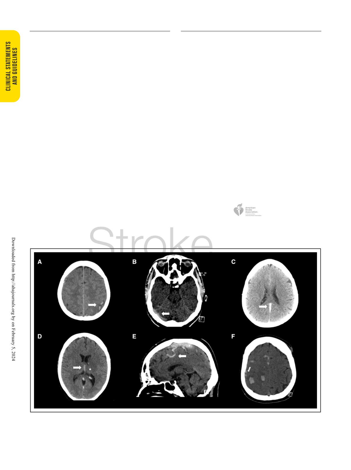
low sensitivity (Figure 2A).36 Bilateral or multifocal hem-
obstruction at the tissue level (venous infarction, edema
orrhages also occur frequently (Figure 2F). In a recent
and hemorrhagic transformation, intracranial hyperten-
meta-analysis of 27 publications with 2812 cases, CT
sion and hydrocephalus) and at the vascular level (dilated
had a sensitivity of 0.79 (95% CI, 0.76-0.82) and speci-
veins).1,34 Common challenges of brain imaging are sum-
ficity of 0.90 (95% CI, 0.89- 0.91),35 lower than previ-
marized in Supplemental Table 2.
ously reported in the 2011 American Heart Association
Direct imaging of thrombus is possible on CT, espe-
statement.1
cially with the increased use of thin-slice CT (Fig-
Thrombi can also be directly observed on conven-
ure 2A-2D).1,35 On noncontrast CT this is observed as
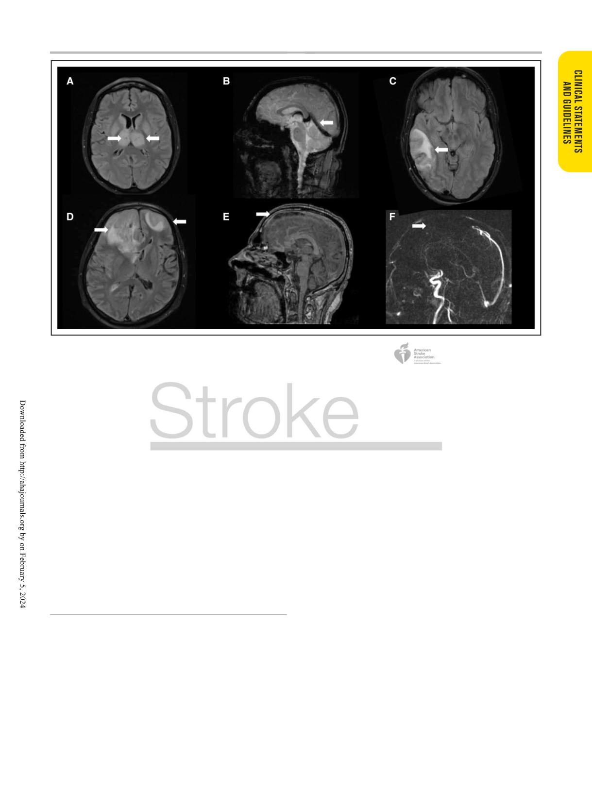
tional MRI sequences (Figure 3).1,37 Because the evolu-
hyperattenuation due to increase of hemoglobin and
tion of a thrombus on MRI is dynamic, changes in the
red blood cells within the thrombus (dense vessel sign;
signal intensity of the thrombus over time are similar
Figure 2B-2E). The traditional cord or string sign, a ser-
to that of a hematoma. As the thrombus ages, oxyhe-
piginous or linear hyperdensity within a vein, or dense
moglobin is converted to deoxyhemoglobin and methe-
triangle sign can be present up to 14 days after onset
moglobin, leading to changes in signal characteristics
of symptoms (Figure 2E). Indirect signs that raise the
on the T1 and T2 sequences. In these early stages, it
suspicion on noncontrast CT (and MRI) include areas
is difficult to diagnose thrombosis because T2 may
of hypodensity not conforming to typical wedge-shaped
be isointense or hypointense, mimicking a normal flow
infarctions or that are not limited to specific arte-
void of a venous sinus. Similarly, time-of-flight magnetic
rial territories or sparing the overlying cortex. Bilateral
resonance venography (MRV) is susceptible to misdiag-
hypodensities may occur when the sagittal sinus or deep
nosis because absent flow is not always corroborated
cerebral veins are involved. Hemorrhages are present
on T1//T2 sequences (Supplemental Table 2). Conse-
in up to 40% of CVT and include areas of hemorrhagic
quently, it is often helpful to corroborate findings with
transformation within regions of hypodensity or frank
gradient-recalled echo, susceptibility-weighted imaging
intracerebral hemorrhage associated with subarachnoid
sequences, or contrast-enhanced MRV.34,37 Thrombosed
or subdural hemorrhages.2,4,16 Isolated subarachnoid
blood creates a blooming artifact on gradient-recalled
Figure 2. Typical findings of cerebral venous thrombosis on noncontrast computed tomography.
A, Left-sided juxtacortical C-shaped hemorrhages. B, Transverse sinus thrombosis. C, Straight sinus thrombosis. D, Internal cerebral vein
thrombosis (arrow) and left thalamic hypodensity (*). E, Cord sign (arrow) and hyperdense sagittal sinus thrombosis (*). F, Multiple small
hemorrhages in same patient as in E. Arrows indicate cord sign.
e4 TBD 2024
Stroke. 2024;55:e00-e00. DOI: 10.1161/STR.0000000000000456
Saposnik et al
Diagnosis and Management of Cerebral Venous Thrombosis
Figure 3. Typical findings of cerebral venous thrombosis on magnetic resonance imaging.
A, Bilateral thalamic hyperintensity (arrows) on fluid-attenuated inversion recovery (FLAIR) in a patient with deep cerebral vein thrombosis. B,
Susceptibility-weighted imaging shows hypointensity of the straight sinus (arrow), vein of Galen, and internal cerebral veins. C, Venous infarction
due to transverse sinus thrombosis with heterogeneous FLAIR hyperintensity (arrow). D, Bilateral FLAIR hyperintensities (arrows) with mass
effect in a patient with superior sagittal sinus thrombosis (arrow), shown in E on a contrast-enhanced T1 sequence and in F absent venous filling
defect (arrow) with phase-contrast magnetic resonance venography.
echo or susceptibility-weighted imaging sequences that
from arachnoid granulations. Several small to moderately
is especially useful in the identification of inconspicuous
sized studies have demonstrated a high sensitivity and
findings (Figure 3B) such as thrombosed cortical veins,
specificity of CTV compared with digital subtraction angi-
where they have a sensitivity and specificity approaching
ography or a consensus reading of other imaging modali-
100%.34,37 Advanced MRI techniques such as T1-based
ties. Compared with MRI, CTV has a lower sensitivity for
black-blood imaging (in which signal from flowing blood
cortical vein thrombosis.37
is suppressed) are promising. MRI is more sensitive than
MRV (Figure 3) can be performed without contrast,
CT in the detection of parenchymal brain lesions second-
with time-of-flight (TOF) or phase-contrast techniques,
ary to venous occlusion such as venous infarctions (Fig-
or with a contrast-enhanced technique (Figure 3E and
ure 3A, 3C, and 3D).38 Radiologically, these lesions cross
3F). The use of gadolinium with contrast-enhanced tech-
arterial vascular territories and may be bilateral.
nique allows direct assessment of luminal filling and
A meta-analysis of 21 studies with 1773 patients
increases the sensitivity of the detection of thrombus
with CVT showed conventional MRI sequences to have a
within the smaller veins.1,37 Both TOF and phase-contrast
sensitivity of 0.82 (95% CI, 0.78-0.85) and specificity of
MRV techniques can be prone to artifact secondary to
0.92 (95% CI, 0.91-0.94).35
complex flow. TOF is, however, still commonly used and is
especially useful in situations that may preclude gadolin-
ium administration such as in pregnant or breastfeeding
CONFIRMING THE DIAGNOSIS OF CVT
patients or in patients with severe renal failure. Compared
CT venography and MRV are the optimal tests to con-
with 3-dimensional TOF, 2-dimensional TOF has higher
firm a diagnosis of CVT. Digital subtraction angiogra-
sensitivity in the setting of slow flow. Phase-contrast MRI
phy is typically used only when invasive treatments are
is used less frequently because defining the velocity of
considered.1
the encoding parameter is not only difficult but opera-
CT venography allows clear depiction of the superfi-
tor dependent and requires longer acquisition times.
cial and deep cerebral venous system. Thrombi present
Contrast-enhanced MRV has a sensitivity and specific-
as filling defects (“empty delta sign” referred to the supe-
ity comparable to that of CTV but allows better charac-
rior sagittal sinus) and can usually be easily distinguished
terization between the low-flow state and hypoplastic
Stroke. 2024;55:e00-e00. DOI: 10.1161/STR.0000000000000456
TBD 2024 e5
Saposnik et al
Diagnosis and Management of Cerebral Venous Thrombosis
sinus (flow gaps).39 Contrast-enhanced techniques and
of 6 months.25 There was no requirement for lead-in par-
gradient-recalled echo or susceptibility-weighted imag-
enteral anticoagulation. The trial excluded individuals with
ing are recommended for diagnosing cortical vein throm-
pregnancy and antiphospholipid antibody syndrome. Nine-
bosis (new evidence since the 2011 American Heart
teen of 26 individuals in the rivaroxaban group received
Association statement).1,34,37
≤4 days of lead-in parenteral anticoagulation.
There were no safety concerns related to early initia-
tion of DOAC. By 6 months, there were 1 recurrent CVT
THERAPEUTIC ADVANCES IN THE
(3.8% [95% CI, 0.1%-19.6%]), 1 symptomatic intrace-
MANAGEMENT OF CVT
rebral hemorrhage (3.8% [95% CI, 0.1%-19.6%]), and 2
clinically relevant nonmajor bleeding events (7.7% [95%
Oral Anticoagulation
CI, 0.9%-25.1%]) in the rivaroxaban group and no VTE
The objectives of anticoagulation therapy in CVT are
recurrence or bleeding events in the control group.
to prevent thrombus growth, to facilitate recanalization,
ACTION-CVT, a large retrospective international study,
and to prevent recurrent VTE events. Previous Ameri-
compared events in 845 consecutive individuals with CVT
can Heart Association/American Stroke Association
who were prescribed VKA versus DOAC as part of their
and European guidelines for the management of CVT
routine clinical care between 2015 and 2020.8 DOACs
recommend the initial use of low-molecular-weight
used included apixaban (67%), rivaroxaban (18%), and
heparin (LMWH) over unfractionated heparin followed
dabigatran (14%) or multiple DOACs (3%). Individuals
by transition to oral vitamin K antagonists (VKAs) for 3
with malignancy, those with antiphospholipid antibody
to 12 months in the context of transient risk factors or
syndrome, and those who were pregnant were excluded.
indefinitely in the context of chronic major risk factors
The study found no significant difference in rates of
for thrombosis or recurrent VTE (Figure 4).1,16,40 LMWH
recurrent VTE (adjusted hazard ratio, 0.94 [95% CI, 0.15-
is favored in the acute treatment of CVT given the more
1.73]), and there was a reduced risk of major hemorrhage
practical administration, more predictable anticoagulation
(adjusted hazard ratio, 0.35 [95% CI, 0.15-0.82]), driven
effect, lower risk of thrombocytopenia, and trends toward
primarily by a lower risk of intracerebral hemorrhage, in
better outcomes in meta-analyses that do not meet the
the DOAC group.8 There were no differences in recanali-
threshold for statistical significance.41,42 The presence of
zation rates between groups in either study.8,47
venous hemorrhage does not constitute a contraindica-
A recent systematic review summarizing 3 randomized
tion for anticoagulation.1,16,43 Whether degree of venous
trials and 16 observational studies comparing DOACs
recanalization should inform duration of anticoagulation
with VKAs found similar rates with both treatments of
remains an area of uncertainty.44-46
recurrent VTE, major hemorrhage, and complete recana-
An emerging body of evidence suggests that direct
lization (42.9% versus 42.3%; relative risk, 0.98 [95% CI,
oral anticoagulants (DOACs), which have demonstrated
0.87-1.11]).48
efficacy and safety compared with VKA for individuals
From the current available evidence, it is reasonable
with deep venous thrombosis and pulmonary embolism,
to transition to DOAC or VKA after a period of lead-in
may also be a reasonable choice for oral anticoagulation
parenteral anticoagulation, but whether 5 to 15 days is
in selected individuals with CVT.
a safer or more effective strategy than shorter periods is
RE-SPECT CVT (A Clinical Trial Comparing Efficacy
not known.47
and Safety of Dabigatran Etexilate With Warfarin in Patients
Additional clinical trials and prospective observational
With Cerebral Venous and Dural Sinus Thrombosis) was an
studies are ongoing (ClinicalTrials.gov NCT03178864,
international prospective clinical trial that randomized 120
NCT04660747). Persistent areas of controversy include
individuals with CVT 1:1 to warfarin VKA with target inter-
timing of initiation with or without lead-in heparin, whether
national normalized ratio of 2.0 to 3.0 or dabigatran 150
acute VTE dosing is initially required, and optimal can-
mg twice daily for 6 months after 5 to 15 days of lead-in
didates for DOAC therapy. DOACs are not suitable in
parenteral anticoagulation.47 The trial excluded individuals
women who are pregnant (both DOAC and warfarin are
with malignancy, central nervous system infection, trauma,
contraindicated; only LMWH is recommended) or breast-
and pregnancy. There were no recurrent VTEs in either
feeding (DOACs are contraindicated; Figure 4). DOACs
group, with 1 (1.7% [95% CI, 0.0%-8.9%]) major hemor-
have also been associated with higher risks of recurrent
rhage (gastrointestinal bleeding) in the dabigatran group
thromboembolic events compared with warfarin in indi-
and 2 (3.3% [95% CI, 0.4%-11.5%], both intracerebral
viduals with antiphospholipid antibody syndrome.49,50 For
hemorrhages) in the warfarin group.
patients with cancer, DOACs were at least noninferior
SECRET (Study of Rivaroxaban in Cerebral Venous
to LMWH in the prevention of VTE. There is limited evi-
Thrombosis) was a phase II trial that randomized 53 par-
dence of the role of antiplatelet agents after discontinu-
ticipants with CVT 1:1 to rivaroxaban 20 mg daily versus
ation of oral anticoagulation in CVT. Studies focused on
standard-of-care anticoagulation (warfarin; target interna-
secondary prevention of VTE showed that aspirin was
tional normalized ratio, 2.0-3.0] or LMWH) for a minimum
more effective than placebo (hazard ratio, 0.68 [95% CI,
e6 TBD 2024
Stroke. 2024;55:e00-e00. DOI: 10.1161/STR.0000000000000456
Saposnik et al
Diagnosis and Management of Cerebral Venous Thrombosis
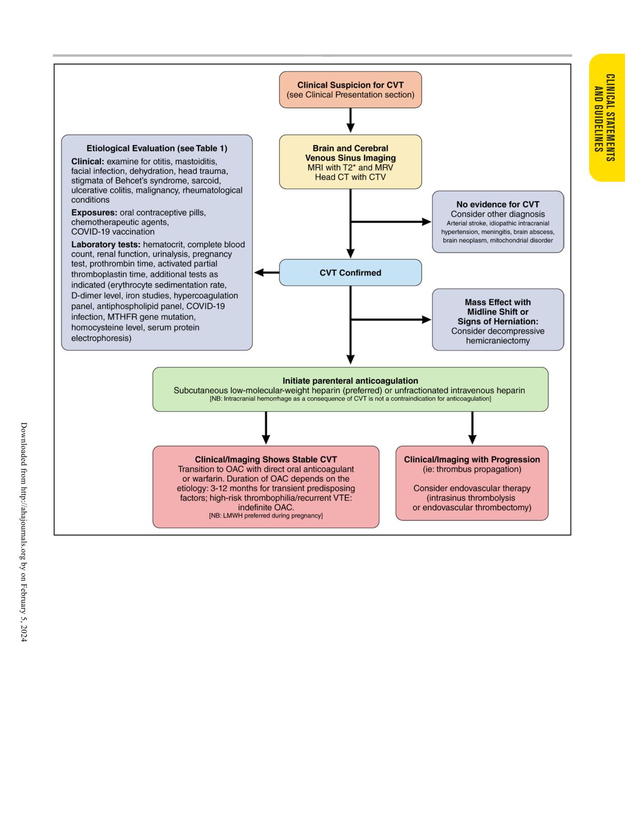
Figure 4. Proposed algorithm for the management of CVT.
This figure summarizes the suggested approach for the diagnosis and management of CVT. CT indicates computed tomography; CTV, computed
tomographic venography; CVT, cerebral venous thrombosis; LMWH, low-molecular-weight heparin; MRI, magnetic resonance imaging; MRV,
magnetic resonance venography; OAC, oral anticoagulant; and VTE, venous thromboembolism.
0.51-0.90]; P=0.008).51 Compared with all comers with
population, is uncertain.52 Several studies in the past
VTE, those with CVT are younger and thus would have
decade that reported the use of mechanical thrombec-
a longer lifetime exposure to aspirin. In addition, the CVT
tomy (either balloon assisted or through aspiration or
literature describing longer-term recurrence risk of VTE
vacuum aspiration systems), intrasinus thrombolysis,
is less robust.31,33 Thus, potential benefits should be care-
combination of mechanical thrombectomy and intra-
fully considered along with individual patient characteris-
sinus thrombolysis, intra-arterial thrombolysis, and intra-
tics in shared decision-making.
sinus stenting provide controversial evidence on safety
and complication rates.43,53 The multicenter, randomized
clinical TO-ACT trial (Thrombolysis or Anticoagulation for
Reperfusion Therapies
CVT) showed that patients with severe CVT did not ben-
Endovascular treatment (EVT) options for the manage-
efit clinically from EVT compared with the patients receiv-
ment of CVT could theoretically offer faster recanaliza-
ing standard anticoagulation therapy.54 Larger studies
tion, although any association with a more favorable
and meta-analyses showed that EVT is associated with
outcome in medical therapy, particularly in an unselected
higher mortality and no evidence of benefit with EVT.55-58
Stroke. 2024;55:e00-e00. DOI: 10.1161/STR.0000000000000456
TBD 2024 e7
Saposnik et al
Diagnosis and Management of Cerebral Venous Thrombosis
Currently, EVT is used as a rescue treatment for
there is deterioration in level of consciousness, more inva-
patients who are experiencing clinical deterioration
sive procedures should be considered and may poten-
or failed or have contraindications to standard therapy
tially be lifesaving (Figure 4). Anticoagulation in the acute
(Figure 4).53 In a systematic review including 10 studies
phase is more controversial in premature and term neo-
comprising 339 patients who underwent EVT for CVT,
nates but is reasonable because there is no evidence of
the authors found a complete and partial postoperative
posttreatment hemorrhage, whereas lack of anticoagula-
recanalization in 90.0% of patients, increasing to 95.2%
tion may lead to thrombus propagation.65,66 CVT in chil-
during the follow-up.59 The complication rate was 10.3%.
dren carries a mortality rate of 3%, and recurrent venous
There is no current evidence to determine which EVT
thrombosis occurs in ≈6%, usually associated with no
technique (eg, stent retriever, microcatheters, aspiration
administration of anticoagulation and lack of venous
catheters, aspiration pump systems) is superior to other
recanalization.64 Since the prior scientific statement,1
therapeutic strategies.
there have been randomized trials on duration of antico-
agulation and the use of DOACs.67 In individuals 4 months
to 20.9 years of age, the Kids-DOTT trial (Multicenter
Decompressive Craniectomy
Evaluation of the Duration of Therapy for Thrombosis in
The evidence on decompressive craniectomy for CVT
Children) compared 6 weeks with 3 months of anticoagu-
remains unchanged since the previous American Heart
lation for provoked VTE (including 59/417; 25% patients
Association statement.1 It should be offered to patients
with CVT). Six weeks of anticoagulation was noninferior
with acute severe CVT and parenchymal lesions with
for recurrent VTE and bleeding events.67
impending herniation as a lifesaving therapeutic approach
In the EINSTEIN-Jr trial (Oral Rivaroxaban in Children
(Figure 4).60 Factors associated with poorer outcomes
With Venous Thrombosis), after initial heparinization, 114
included age >50 years, midline shift >10 mm, and total
children with confirmed CVT were randomized (2:1) to 3
effacement of basal cisterns.61 There are no randomized
months of rivaroxaban or standard anticoagulation (con-
controlled trials of this surgical approach in the litera-
tinuing heparin or switching to oral VKAs).68 The primary
ture. A systematic review and meta-analysis including 51
efficacy outcome was symptomatic recurrent VTE, and
studies comprising 483 patients with CVT showed that
the principal safety outcome was major or clinically rel-
surgery within 48 hours of admission may decrease mor-
evant nonmajor bleeding. With 100% follow-up, none of
tality (odds ratio [OR], 0.26 [95% CI, 0.10-0.69]) and
the 73 children treated with rivaroxaban compared with 1
result in improved functional outcomes.62
of the 41 children treated with standard anticoagulation
had symptomatic recurrent VTE (absolute difference,
2.4% [95% CI, -2.6% to 13.5%]). Five patients on rivar-
CVT IN SPECIAL POPULATIONS
oxaban had nonmajor and noncerebral bleeding, whereas
1 patient on standard anticoagulation had a major sub-
Pediatric Population
dural bleed. Complete or partial recanalization occurred
CVT is more common in neonates (6.4/100 000) than
in 18 (25%) and 39 (53%) patients on rivaroxaban and
in children and adolescents.63 The key to successful
6 (15%) and 24 (59%) patients on standard antico-
management is to consider the diagnosis early in acute
agulation. No children died by the end of the 3-month
presentations with headache, seizures, focal neurological
study treatment period. Focal neurological deficits were
deficits, or coma and in typical situations such as sepsis
observed in 5 (6.8%) and 3 (7.3%) children in the rivar-
(including mastoiditis, Lemierre syndrome, COVID-19,
oxaban and standard anticoagulation group, respectively,
and meningitis), head trauma (including abuse), hypoxia,
at the end of the study. However, other long-term stud-
and dehydration. Awareness is especially important in
ies suggest that despite treatment 1 in 4 children may
children with preexisting conditions (congenital heart dis-
develop late epilepsy,69 infantile spasms after neonatal
ease and its surgical treatment, cancer and its treatment,
CVT, cognitive impairment, or intracranial hypertension.70
anemia due to iron deficiency or hemoglobinopathies,
and inflammatory conditions, eg, nephrotic syndrome
CVT During Pregnancy and Puerperium
and inflammatory bowel disease). Emergency imaging to
diagnose or exclude dural or cortical venous thrombosis
Pregnancy induces changes in the coagulation system
with or without parenchymal involvement or hemorrhage
that persist into the puerperium and result in a hyper-
typically requires anesthesia. Initial blood work may reveal
coagulable state, increasing the risk of CVT. Incidence
low platelets associated with platelet factor 4 mutations
estimates during pregnancy and the puerperium range
or high platelets and high hemoglobin associated with
from 1 in 2500 deliveries to 1 in 10 000 deliveries in
JAK2 mutations, as well as indices of iron deficiency with
Western countries, and ORs range from 1.3 to 13.0. The
or without anemia.63,64
greatest risk periods for CVT include the third trimester
The management of acute CVT in children typically
and the first 6 postpartum weeks. Approximately 80%
involves LMWH or unfractionated heparin. As for adults, if
of pregnancy-related CVT cases occur after delivery.12 A
e8 TBD 2024
Stroke. 2024;55:e00-e00. DOI: 10.1161/STR.0000000000000456
Saposnik et al
Diagnosis and Management of Cerebral Venous Thrombosis
case-control study found that the risk is increased only in
poor prognosis, with mortality rates ranging from 39% to
the puerperium (OR, 10.6 [95% CI, 5.6-20.0]), whereas
61% in initial cohort studies.80,82,83 A study on the global
there was no statistically significant increased risk of CVT
impact of COVID-19 that included 2313 CVT admissions
during pregnancy (OR, 1.2 [95% CI, 0.6-2.3]).10 Cesar-
across 1 prepandemic year (2019) and 1 pandemic year
ean delivery appears to be associated with a higher risk
(2020) found 26 cases of VITT, resulting in 6 deaths. A
after adjustment for age, vascular risk factors, presence
diagnosis of COVID-19 infection was present in 7.6% of
of infections, hospital type, and location (OR, 3.10 [95%
all CVT admissions.19
CI, 2.26-4.24]).10 Overall, studies published since the
A pharmacovigilance study comprising
>1.7 mil-
prior scientific statement1 have found that the prognosis
lion adverse reactions showed a disproportionately
for women with pregnancy-related CVT is better than or
lower incident risk of CVT after vaccination with mRNA
the same as that for patients with CVT in general.71
SARS-CoV-2 vaccines compared with adenovirus-based
VKAs, including warfarin, are associated with fetal
SARS-CoV-2 vaccines (1-5/10 000 for BNT162b2 and
embryopathy and bleeding in the fetus and neonate and
mRNA-1273 versus 13/10 000 for ChAdOx1 nCoV-19
thus are contraindicated in pregnancy. Therefore, antico-
vaccine).84 In addition, there is no evidence of VITT after
agulation for CVT during pregnancy and early in the puer-
mRNA vaccines.76,84
perium consists of LMWH in the majority of women.12,72,73
In cases of suspected VITT, laboratory testing for
There is limited evidence on endovascular therapies in
platelet factor 4 antibodies is recommended. Despite
this population. As in nonpregnant women, thrombolysis
the lack of evidence, given the similarity to autoimmune
and thrombectomy are reserved for patients with neu-
heparin-induced thrombocytopenia, avoidance of hepa-
rological deterioration or propagation of the thrombus
rin products, intravenous immunoglobulin 1 g/kg body
despite medical therapy (Figure 4).
weight daily for 2 days, and administration of steroids
have been advised. Platelet transfusions are not rec-
ommended.85 Nonheparin parenteral anticoagulants
Future Pregnancies and Recurrence
(argatroban, fondaparinux) are typically used first, with
Women with a history of VTE appear to have an increased
transition to an oral anticoagulant (ie, DOAC) once there
risk of thrombotic events (ie, deep venous thrombosis,
is full platelet count recovery.80
pulmonary embolism) in future pregnancies.64 A system-
atic review comprising 17 studies and 393 pregnan-
cies found a recurrence rate of 8 per 1000 pregnancies
KEY POINTS FOR CLINICAL PRACTICE
(95% CI, 3-22). The rate of noncerebral VTEs was 22
• CVT requires a high level of suspicion among patients
per 1000 pregnancies (95% CI, 11-43).74 There was a
presenting with common symptoms and known pre-
trend toward a lower rate of recurrent thrombotic events
disposing conditions (pregnancy, puerperium, use of
in women who used antithrombotic prophylaxis.74
oral contraceptives, thrombophilia) or demographic
According to the available evidence, CVT is not a
factors (young women).
contraindication for future pregnancies.12,72,75 Consider-
• New predisposing conditions (obesity, COVID-19,
ing the additional risk that pregnancy confers to women
vaccine-induced thrombocytopenia)2,15,18 were identi-
fied since our previous report.1 (New)
with a history of CVT, prophylaxis with LMWH during
• MRI/MRV is the recommended noninvasive study of
future pregnancies and the postpartum period is prob-
the cerebral venous system to confirm the diagnosis.
ably beneficial.75
CT/CTV is a reasonable alternative in centers with
limited resources or if the pretest probability is low.
• Contrast-enhanced MRV, gradient-recalled echo, or
VITT and CVT
susceptibility-weighted imaging sequences are the
In 2021, reports from Europe and the United States
recommended techniques for the diagnosis of corti-
described thrombocytopenia and CVT after vaccination
cal venous thrombosis. (New)
with the ChAdOx1 nCoV-19 vaccine (AstraZeneca) and
• The mainstream initial treatment of CVT includes par-
the Ad26.COV2.S adenovirus-based SARS-CoV-2 vac-
enteral heparin followed by transition to oral VKAs for
cine (Janssen).76-78 The age range of affected patients
3 to 12 months, depending on the underlying cause,
or indefinitely in the presence of thrombophilia or
was 18 to 77 years; most were women. Symptoms began
recurrent VTE (Figure 4).
5 to 24 days after vaccination.76 Headache was the most
• DOACs appear to be a safe and effective alternative
common presenting feature. All patients had throm-
option to VKAs according to open-label retrospective
bocytopenia. In the United Kingdom, 23 patients with
and prospective randomized studies. (New)
antibodies to platelet factor 4 after ChAdOx1 nCoV-19
• The strategy of identifying venous recanalization in
vaccination were described. It is believed that DNA from
subsequent CTV or MRV to guide the duration of anti-
the adenovirus-infected cells bonded to platelet factor
coagulation remains uncertain. (New)
4 and triggered the production of autoantibodies.79,80
• Given the lack of controlled studies (and poorer out-
Although CVT in VITT is a rare condition,81 it carries a
comes in meta-analyses), endovascular therapies
Stroke. 2024;55:e00-e00. DOI: 10.1161/STR.0000000000000456
TBD 2024 e9

Saposnik et al
Diagnosis and Management of Cerebral Venous Thrombosis
are reserved for patients with evidence of thrombus
ARTICLE INFORMATION
propagation, for individuals with neurological dete-
The American Heart Association makes every effort to avoid any actual or poten-
rioration despite medical therapy, or for those with
tial conflicts of interest that may arise as a result of an outside relationship or a
contraindications to anticoagulation (Figure 4).
personal, professional, or business interest of a member of the writing panel. Spe-
cifically, all members of the writing group are required to complete and submit a
(New)
Disclosure Questionnaire showing all such relationships that might be perceived
• Despite the low level of evidence, decompressive
as real or potential conflicts of interest.
surgery is a lifesaving procedure that may result in
This statement was approved by the American Heart Association Science Ad-
improved functional outcomes among patients with
visory and Coordinating Committee on October 11, 2023, and the American Heart
Association Executive Committee on December 4, 2023. A copy of the document
advanced clinical signs of herniation. (New)
is available at https://professional.heart.org/statements by using either “Search
• For women with CVT during pregnancy, LMWH in full
for Guidelines & Statements” or the “Browse by Topic” area. To purchase additional
anticoagulant doses should be continued throughout
reprints, call 215-356-2721 or email Meredith.Edelman@wolterskluwer.com
pregnancy, and LMWH or VKA with a target interna-
The American Heart Association requests that this document be cited as
tional normalized ratio of 2.0 to 3.0 should be con-
follows: Saposnik G, Bushnell C, Coutinho JM, Field TS, Furie KL, Galadanci N,
Kam W, Kirkham FC, McNair ND, Singhal AB, Thijs V, Yang VXD; on behalf of the
tinued for at least 6 weeks postpartum (for a total
American Heart Association Stroke Council; Council on Cardiopulmonary, Criti-
minimum duration of therapy of 3 months).
cal Care, Perioperative and Resuscitation; Council on Cardiovascular and Stroke
• It is reasonable to advise women with a history of
Nursing; and Council on Hypertension. Diagnosis and management of cerebral
CVT that future pregnancy is not contraindicated.
venous thrombosis: a scientific statement from the American Heart Association.
Stroke. 2024;55:e•••-e•••. doi: 10.1161/STR.0000000000000456
Prophylaxis with LMWH during future pregnancies
The expert peer review of AHA-commissioned documents (eg, scientific
and the postpartum period is usually recommended.
statements, clinical practice guidelines, systematic reviews) is conducted by the
• CVT in the pediatric population is more common in
AHA Office of Science Operations. For more on AHA statements and guidelines
neonates than children, usually among those exposed
development, visit https://professional.heart.org/statements. Select the “Guide-
to infections, dehydration, iron deficiency, anemia, or
lines & Statements” drop-down menu, then click “Publication Development.”
Permissions: Multiple copies, modification, alteration, enhancement, and dis-
head trauma. Parenteral anticoagulation is also the
tribution of this document are not permitted without the express permission of the
first-line treatment, followed by LMWH, VKA, or rivar-
American Heart Association. Instructions for obtaining permission are located at
oxaban for at least 6 weeks.
• VITT and CVT may occur (rarely) days or a few weeks
Form” appears in the second paragraph (https://www.heart.org/en/about-us/
statements-and-policies/copyright-request-form).
after an individual receives adenovirus-based SARS-
CoV-2 vaccines, usually presenting with new onset
Acknowledgments
of headaches and thrombocytopenia; it requires the
The authors appreciate the support of Dr Gokce H. Majernik (vascular fellow at
expert management of a hematologist and multidisci-
the London Health Sciences Center, London, Ontario, Canada) for reviewing the
plinary team. (New)
section on endovascular therapy in CVT.
Disclosures
Writing Group Disclosures
Writing
Other
Speakers’
Consultant/
group
research
bureau/
Expert
Ownership
advisory
member
Employment
Research grant
support
honoraria
witness
interest
board
Other
Gustavo
St Michael’s Hos-
None
None
None
None
None
None
None
Saposnik
pital, University of
Toronto (Canada)
Cheryl
Wake Forest
None
None
None
None
None
None
None
Bushnell
School of
Medicine
Jonathan
Amsterdam
Bayer*; AstraZeneca*; Dutch
None
None
None
TrianecT*
None
None
M. Coutinho
UMC, University
Heart Foundation (nonprofit
of Amsterdam
organization)*; Dutch Thrombosis
(Netherlands)
Foundation (nonprofit
organization)*; ZonMw (nonprofit
government organization)*; All fees
paid to his employer for all.
Thalia S.
University of
Canadian Institutes of Health
None
None
Canadian
DESTINE
None
None
Field
British Columbia
Research (research grant to her
Medical
Health*
Centre for Brain
institution)†; Heart and Stroke
Protective
Health (Canada)
Foundation of Canada (research
Association*
grant to her institution)†;
Bayer Canada (in-kind study
medication)†
Karen L
Rhode Island
None
None
None
None
None
None
None
Furie
Hospital
(Continued )
e10 TBD 2024
Stroke. 2024;55:e00-e00. DOI: 10.1161/STR.0000000000000456
Saposnik et al
Diagnosis and Management of Cerebral Venous Thrombosis
Writing Group Disclosures Continued
Writing
Other
Speakers’
Consultant/
group
research
bureau/
Expert
Ownership
advisory
member
Employment
Research grant
support
honoraria
witness
interest
board
Other
Najibah
University of
None
None
None
None
None
None
None
Galadanci
Alabama at
Birmingham
Wayneho
Duke University
None
None
None
None
None
None
None
Kam
Hospital; UNC
Health Rex
Comprehensive
Stroke Center
Fenella J.
UCL GOSH
Action Medical Research (RCT
None
None
None
None
Pfizer†
UCL
Kirkham
Institute of Child
of Montelukast in SCD with
(professor
Health (United
processing speed end point)†
of pediatric
Kingdom)
neurology)†
Norma D.
Retired Nursing
None
None
None
None
None
None
None
McNair
Aneesh B.
Massachusetts
MGH Fireman Vascular Center
None
None
MedicoLegal
None
None
None
Singhal
General Hospital
(SPARK Award)*
expert
witness*
Vincent
Florey Neurology
None
None
Boehringer
None
None
None
None
Thijs
(Australia)
Ingelheim*
Victor X. D.
Sunnybrook
None
None
None
None
None
None
None
Yang
Health Sciences
Center (Canada)
This table represents the relationships of writing group members that may be perceived as actual or reasonably perceived conflicts of interest as reported on the
Disclosure Questionnaire, which all members of the writing group are required to complete and submit. A relationship is considered to be “significant” if (a) the person
receives $5000 or more during any 12-month period, or 5% or more of the person’s gross income; or (b) the person owns 5% or more of the voting stock or share of the
entity, or owns $5000 or more of the fair market value of the entity. A relationship is considered to be “modest” if it is less than “significant” under the preceding definition.
*Modest.
†Significant.
Reviewer Disclosures
Other
Speakers’
Consultant/
Research
research
bureau/
Expert
Ownership
advisory
Reviewer
Employment
grant
support
honoraria
witness
interest
board
Other
Ekaterina
University of Alabama
None
None
None
None
None
None
None
Bakradze
at Birmingham
José Ferro
Faculdade
None
None
None
None
None
None
None
de Medicina,
Universidade de
Lisboa (Portugal)
Matthew
Massachusetts
None
None
None
None
None
None
None
Frosch
General Hospital
Hans-
Vivantes Klinikum
None
None
None
None
None
None
Medical guidelines (I am member of
Christian
im Friedrichshain
the writing group for guidelines for
Koennecke
(Germany)
cerebral venous thrombosis of the
Deutsche Gesellschaft für Neurologie
[German Neurological Society])*
Thanh
Boston University
None
None
None
None
None
Idorsia*
None
Nguyen
Chobanian and
Avedisian School of
Medicine
Brian Silver
UMass Memorial
None
None
None
None
None
None
None
Medical Center
Kori
Massachusetts
None
None
None
None
None
None
None
Zachrison
General Hospital
This table represents the relationships of reviewers that may be perceived as actual or reasonably perceived conflicts of interest as reported on the Disclosure Ques-
tionnaire, which all reviewers are required to complete and submit. A relationship is considered to be “significant” if (a) the person receives $5000 or more during any
12-month period, or 5% or more of the person’s gross income; or (b) the person owns 5% or more of the voting stock or share of the entity, or owns $5000 or more of
the fair market value of the entity. A relationship is considered to be “modest” if it is less than “significant” under the preceding definition.
*Modest.
Stroke. 2024;55:e00-e00. DOI: 10.1161/STR.0000000000000456
TBD 2024 e11
Saposnik et al
Diagnosis and Management of Cerebral Venous Thrombosis
REFERENCES
19.
Nguyen TN, Qureshi MM, Klein P, Yamagami H, Abdalkader M, Mikulik R,
Sathya A, Mansour OY, Czlonkowska A, Lo H, et al. Global impact of the
1.
Saposnik G, Barinagarrementeria F, Brown RD Jr, Bushnell CD, Cucchiara B,
COVID-19 pandemic on cerebral venous thrombosis and mortality. J Stroke.
Cushman M, deVeber G, Ferro JM, Tsai FY; on behalf of the American
2022;24:256-265. doi: 10.5853/jos.2022.00752
Heart Association Stroke Council and the Council on Epidemiology and
20.
Liu K, Pei L, Gao Y, Zhao L, Fang H, Bunda B, Fisher L, Wang Y, Li S, Li Y,
Prevention. Diagnosis and management of cerebral venous thrombosis:
et al. Dehydration status predicts short-term and long-term outcomes in
a statement for healthcare professionals from the American Heart Asso-
patients with cerebral venous thrombosis. Neurocrit Care. 2019;30:478-
ciation/American Stroke Association. Stroke. 2011;42:1158-1192. doi:
483. doi: 10.1007/s12028-018-0628-3
10.1161/STR.0b013e31820a8364
21.
Shabo E, Wach J, Hamed M, Guresir A, Weinhold L, Vatter H,
2.
Alet M, Ciardi C, Alemán A, Bandeo L, Bonardo P, Cea C,
Guresir E. Asymptomatic postoperative cerebral venous sinus throm-
Cirio J, Cossio J, Cuculic M, Esnaola MM, et al. Cerebral venous throm-
bosis after posterior fossa tumor surgery: incidence, risk factors,
bosis in Argentina: clinical presentation, predisposing factors, outcomes
and therapeutic options. Neurosurgery.
2023;92:1171-1176. doi:
and literature review. J Stroke Cerebrovasc Dis. 2020;29:105145. doi:
10.1227/neu.0000000000002340
10.1016/j.jstrokecerebrovasdis.2020.105145
22.
Hiltunen S, Putaala J, Haapaniemi E, Tatlisumak T. Long-term outcome
3.
Maali L, Khan S, Qeadan F, Ismail M, Ramaswamy D, Hedna VS. Cerebral
after cerebral venous thrombosis: analysis of functional and vocational out-
venous thrombosis: continental disparities. Neurol Sci. 2017;38:1963-
come, residual symptoms, and adverse events in 161 patients. J Neurol.
1968. doi: 10.1007/s10072-017-3082-7
2016;263:477-484. doi: 10.1007/s00415-015-7996-9
4.
Ferro JM, Canhao P, Stam J, Bousser MG, Barinagarrementeria F; ISCVT
23.
Koopman K, Uyttenboogaart M, Vroomen PC, van der Meer J, De Keyser J,
Investigators. Prognosis of cerebral vein and dural sinus thrombosis: results of
Luijckx GJ. Long-term sequelae after cerebral venous thrombosis in func-
the International Study on Cerebral Vein and Dural Sinus Thrombosis (ISCVT).
tionally independent patients. J Stroke Cerebrovasc Dis. 2009;18:198-202.
Stroke. 2004;35:664-670. doi: 10.1161/01.STR.0000117571.76197.26
doi: 10.1016/j.jstrokecerebrovasdis.2008.10.004
5.
Klein P, Shu L, Nguyen TN, Siegler JE, Omran SS, Simpkins AN, Heldner M,
24.
Preter M, Tzourio C, Ameri A, Bousser MG. Long-term prognosis in cerebral
Havenon A, Aparicio HJ, Abdalkader M, et al. Outcome prediction in cerebral
venous thrombosis: follow-up of 77 patients. Stroke. 1996;27:243-246.
venous thrombosis: the IN-REvASC Score. J Stroke. 2022;24:404-416.
doi: 10.1161/01.str.27.2.243
doi: 10.5853/jos.2022.01606
25.
Field T, Dizonno V, Almekhlafi MA, Bala F, Alhabli I, Wong H, Norena M,
6.
Martinelli I, Bucciarelli P, Passamonti SM, Battaglioli T, Previtali E,
Villaluna MK, King-Azote P, Ratnaweera N, et al; SECRET Investigators. Study
Mannucci PM. Long-term evaluation of the risk of recurrence after cere-
of Rivaroxaban in Cerebral Venous Thrombosis: a randomized controlled
bral sinus-venous thrombosis. Circulation.
2010;121:2740-2746. doi:
feasibility trial comparing anticoagulation with rivaroxaban to standard-of-
10.1161/CIRCULATIONAHA.109.927046
care in symptomatic cerebral venous thrombosis. Stroke. 2023;54:2724-
7.
Duman T, Uluduz D, Midi I, Bektas H, Kablan Y, Goksel BK, Milanlioglu A,
2736. doi: 10.1161/STROKEAHA.123.044113
Necioglu Orken D, Aluclu U. A multicenter study of 1144 patients with
26.
Liu L, Jiang H, Wei H, Zhou Y, Wu Y, Zhang K, Duan J, Meng R, Zhou C,
cerebral venous thrombosis: the VENOST study. J Stroke Cerebrovasc Dis.
Ji X. Risk factors of impaired employability after cerebral venous thrombo-
2017;26:1848-1857. doi: 10.1016/j.jstrokecerebrovasdis.2017.04.020
sis. CNS Neurosci Ther. 2023;29:1086-1093. doi: 10.1111/cns.14083
8.
Yaghi S, Shu L, Bakradze E, Salehi Omran S, Giles JA, Amar JY, Henninger N,
27.
Gasparini S, Neri S, Brigo F, Cianci V, Mammì A, Pascarella A, Manzo L,
Elnazeir M, Liberman AL, Moncrieffe K, et al. Direct oral anticoagulants
Benna P, Striano P, Martino T, et al. Late epileptic seizures following cere-
versus warfarin in the treatment of cerebral venous thrombosis (ACTION-
bral venous thrombosis: a systematic review and meta-analysis. Neurol Sci.
CVT): a multicenter international study. Stroke. 2022;53:728-738. doi:
2022;43:5229-5236. doi: 10.1007/s10072-022-06148-y
10.1161/STROKEAHA.121.037541
28.
Sanchez van Kammen M, Lindgren E, Silvis SM, Hiltunen S, Heldner MR,
9.
Lindgren E, Silvis SM, Hiltunen S, Heldner MR, Serrano F, de Scisco M,
Serrano F, Zelano J, Zuurbier SM, Mansour M, Aguiar de Sousa D, et al. Late
Zelano J, Zuurbier SM, Sanchez van Kammen M, Mansour M, et al.
seizures in cerebral venous thrombosis. Neurology. 2020;95:e1716-e1723.
Acute symptomatic seizures in cerebral venous thrombosis. Neurology.
doi: 10.1212/WNL.0000000000010576
2020;95:e1706-e1715. doi: 10.1212/WNL.0000000000010577
29.
Lindgren E, Rentzos A, Hiltunen S, Serrano F, Heldner MR, Zuurbier SM,
10.
Silvis SM, Lindgren E, Hiltunen S, Devasagayam S, Scheres LJ, Jood K,
Silvis SM, Mansour M, Allingham W, Punter MNM, et al. Dural arteriovenous
Zuurbier SM, Kleinig TJ, Silver FL, Mandell DM, et al. Postpartum period is a
fistulas in cerebral venous thrombosis: data from the International Cere-
risk factor for cerebral venous thrombosis. Stroke. 2019;50:501-503. doi:
bral Venous Thrombosis Consortium. Eur J Neurol. 2022;29:761-770. doi:
10.1161/STROKEAHA.118.023017
10.1111/ene.15192
11.
Amoozegar F, Ronksley PE, Sauve R, Menon BK. Hormonal contraceptives
30.
Ferro JM, Coutinho JM, Jansen O, Bendszus M, Dentali F, Kobayashi A,
and cerebral venous thrombosis risk: a systematic review and meta-analysis.
van der Veen B, Miede C, Caria J, Huisman H, et al. Dural arteriovenous
Front Neurol. 2015;6:7. doi: 10.3389/fneur.2015.00007
fistulae after cerebral venous thrombosis. Stroke. 2020;51:3344-3347. doi:
12.
Khan M, Arauz A, Uluduz D, Barboza MA, Duman T, Cano-Nigenda V, Awan S,
10.1161/STROKEAHA.120.031235
Wasay M; VENOST Study Group. Predictors of mortality and functional out-
31.
Palazzo P, Agius P, Ingrand P, Ciron J, Lamy M, Berthomet A,
come in pregnancy and puerperium-related cerebral venous thrombosis.
Cantagrel P, Neau JP. Venous thrombotic recurrence after cerebral venous
Cerebrovasc Dis. 2023;52:393-400. doi: 10.1159/000527155
thrombosis: a long-term follow-up study. Stroke. 2017;48:321-326. doi:
13.
Kashkoush AI, Ma H, Agarwal N, Panczykowski D, Tonetti D, Weiner GM,
10.1161/strokeaha.116.015294
Ares W, Kenmuir C, Jadhav A, Jovin T, et al. Cerebral venous sinus thrombo-
sis in pregnancy and puerperium: a pooled, systematic review. J Clin Neuro-
32.
Shu L, Bakradze E, Omran SS, Giles J, Amar J, Henninger N,
sci. 2017;39:9-15. doi: 10.1016/j.jocn.2017.02.046
Elnazeir M, Liberman A, Moncrieffe K, Rotblat J, et al. Predictors of
14.
Getahun D, Nash R, Flanders WD, Baird TC, Becerra-Culqui TA, Cromwell L,
recurrent venous thrombosis after cerebral venous thrombosis: analy-
Hunkeler E, Lash TL, Millman A, Quinn VP, et al. Cross-sex hormones and
sis of the ACTION-CVT study. Neurology. 2022;99:e2368-e2377. doi:
acute cardiovascular events in transgender persons: a cohort study. Ann
10.1212/wnl.0000000000201122
Intern Med. 2018;169:205-213. doi: 10.7326/m17-2785
33.
Skajaa N, Vandenbroucke JP, Simonsen CZ, Sørensen HT, Adelborg K.
15.
Zuurbier SM, Arnold M, Middeldorp S, Broeg-Morvay A, Silvis SM,
Thromboembolic events, bleeding, and mortality in patients with cerebral
Heldner MR, Meisterernst J, Nemeth B, Meulendijks ER, Stam J, et al. Risk of
venous thrombosis: a nationwide cohort study. Blood Adv. 2023;7:2070-
cerebral venous thrombosis in obese women. JAMA Neurol. 2016;73:579-
2081. doi: 10.1182/bloodadvances.2022008622
584. doi: 10.1001/jamaneurol.2016.0001
34.
Dmytriw AA, Song JSA, Yu E, Poon CS. Cerebral venous thrombosis: state
16.
Ferro JM, Bousser MG, Canhão P, Coutinho JM, Crassard I, Dentali F,
of the art diagnosis and management. Neuroradiology. 2018;60:669-685.
di Minno M, Maino A, Martinelli I, Masuhr F, et al. European Stroke Organiza-
doi: 10.1007/s00234-018-2032-2
tion guideline for the diagnosis and treatment of cerebral venous thrombo-
35.
Xu W, Gao L, Li T, Ramdoyal ND, Zhang J, Shao A. The performance of
sis. Eur J Neurol. 2017;24:1203-1213. doi: 10.1111/ene.13381
CT versus MRI in the differential diagnosis of cerebral venous thrombosis.
17.
King A, Doyle KM. Implications of COVID-19 to stroke medicine: an epi-
Thromb Haemost. 2018;118:1067-1077. doi: 10.1055/s-0038-1642636
demiological and pathophysiological perspective. Curr Vasc Pharmacol.
36.
Coutinho JM, van den Berg R, Zuurbier SM, VanBavel E, Troost D, Majoie CB,
2022;20:333-340. doi: 10.2174/1570161120666220428101337
Stam J. Small juxtacortical hemorrhages in cerebral venous thrombosis. Ann
18.
Simonetto M, Wechsler PM, Merkler AE. Stroke treatment in the era of
Neurol. 2014;75:908-916. doi: 10.1002/ana.24180
COVID-19: a review. Curr Treat Options Neurol. 2022;24:155-171. doi:
37.
van Dam LF, van Walderveen MAA, Kroft LJM, Kruyt ND, Wermer MJH,
10.1007/s11940-022-00713-8
van Osch MJP, Huisman MV, Klok FA. Current imaging modalities for
e12 TBD 2024
Stroke. 2024;55:e00-e00. DOI: 10.1161/STR.0000000000000456
Saposnik et al
Diagnosis and Management of Cerebral Venous Thrombosis
diagnosing cerebral vein thrombosis: a critical review. Thromb Res.
cerebral venous thrombosis: the TO-ACT randomized clinical trial. JAMA
2020;189:132-139. doi: 10.1016/j.thromres.2020.03.011
Neurol. 2020;77:966-973. doi: 10.1001/jamaneurol.2020.1022
38.
Alajmi E, Zung J, Duquet-Armand M, Coutinho JM, Mandell DM. Prevalence
55.
Siddiqui FM, Weber MW, Dandapat S, Scaife S, Buhnerkempe M,
of venous infarction in patients with cerebral venous thrombosis: baseline
Ortega-Gutierrez S, Aksan N, Elias A, Coutinho JM. Endovascular thrombol-
diffusion-weighted MRI and follow-up MRI. Stroke. 2023;54:1808-1814.
ysis or thrombectomy for cerebral venous thrombosis: study of Nationwide
doi: 10.1161/STROKEAHA.122.042336
Inpatient Sample 2004-2014. J Stroke Cerebrovasc Dis. 2019;28:1440-
39.
Pai V, Khan I, Sitoh YY, Purohit B. Pearls and pitfalls in the magnetic resonance
1447. doi: 10.1016/j.jstrokecerebrovasdis.2019.03.025
diagnosis of dural sinus thrombosis: a comprehensive guide for the trainee
56.
Xu Z, Li X, Feng D, Wang T, Xu X, Deng R, Zhou X, Chen G. Endovas-
radiologist. J Clin Imaging Sci. 2020;10:77. doi: 10.25259/jcis_187_2020
cular therapy versus anticoagulation for treatment of cerebral venous
40.
Einhäupl K, Stam J, Bousser MG, De Bruijn SF, Ferro JM, Martinelli I, Masuhr F;
sinus thrombosis: a meta-analysis. Neurologist.
2021;27:69-73. doi:
European Federation of Neurological Societies. EFNS guideline on the
10.1097/NRL.0000000000000369
treatment of cerebral venous and sinus thrombosis in adult patients. Eur J
57.
Naik A, Smith E, Dharnipragada R, Catapano JS, Cramer SW, Johnson R,
Neurol. 2010;17:1229-1235. doi: 10.1111/j.1468-1331.2010.03011.x
Khanam R, Hassaneen W, Lawton MT, Arnold PM. Endovascular and medi-
41.
Misra UK, Kalita J, Chandra S, Kumar B, Bansal V. Low molecular weight
cal management of cerebral venous thrombosis: a systematic review and
heparin versus unfractionated heparin in cerebral venous sinus thrombo-
network meta-analysis. World Neurosurg.
2022;165:e197-e205. doi:
sis: a randomized controlled trial. Eur J Neurol. 2012;19:1030-1036. doi:
10.1016/j.wneu.2022.05.142
10.1111/j.1468-1331.2012.03690.x
58.
Siegler JE, Shu L, Yaghi S, Salehi Omran S, Elnazeir M, Bakradze E,
42.
Erkens PM, Prins MH. Fixed dose subcutaneous low molecular weight
Psychogios M, De Marchis GM, Yu S, Klein P, et al. Endovascular therapy
heparins versus adjusted dose unfractionated heparin for venous throm-
for cerebral vein thrombosis: a propensity-matched analysis of antico-
boembolism. Cochrane Database Syst Rev.
2010:CD001100. doi:
agulation in the treatment of cerebral venous thrombosis. Neurosurgery.
10.1002/14651858.CD001100.pub3
2022;91:749-755. doi: 10.1227/neu.0000000000002098
43.
Fan Y, Yu J, Chen H, Zhang J, Duan J, Mo D, Zhu W, Wang B, Ouyang F,
59.
Paybast S, Mohamadian R, Emami A, Jameie M, Shahrab F,
Chen Y, et al. Chinese Stroke Association guidelines for clinical manage-
Zamani F, Sharifipour E. Safety and efficacy of endovascular thromboly-
ment of cerebrovascular disorders: executive summary and 2019 update
sis in patients with acute cerebral venous sinus thrombosis: a system-
of clinical management of cerebral venous sinus thrombosis. Stroke Vasc
atic review [published online December 5, 2022]. Interv Neuroradiol.
Neurol. 2020;5:152-158. doi: 10.1136/svn-2020-000358
doi:
10.1177/15910199221143418. https://journals.sagepub.com/
44.
Aguiar de Sousa D, Lucas Neto L, Arauz A, Sousa AL, Gabriel D, Correia M,
doi/10.1177/15910199221143418?url_ver=Z39.88-2003&rfr_
Gil-Gouveia R, Penas S, Carvalho Dias M, Correia MA, et al. Early recanaliza-
id=ori:rid:crossref.org&rfr_dat=cr_pub%20%200pubmed
tion in patients with cerebral venous thrombosis treated with anticoagulation.
60.
Ferro JM, Bousser MG, Canhao P, Coutinho JM, Crassard I, Dentali F,
Stroke. 2020;51:1174-1181. doi: 10.1161/STROKEAHA.119.028532
di Minno M, Maino A, Martinelli I, Masuhr F, et al. European Stroke Organiza-
45.
Ferro JM, Bendszus M, Jansen O, Coutinho JM, Dentali F, Kobayashi A,
tion guideline for the diagnosis and treatment of cerebral venous thrombo-
Aguiar de Sousa D, Neto LL, Miede C, Caria J, et al. Recanalization after
sis. Eur Stroke J. 2017;2:195-221. doi: 10.1177/2396987317719364
cerebral venous thrombosis: a randomized controlled trial of the safety
61.
Mahale R, Mehta A, Varma RG, Hegde AS, Acharya PT, Srinivasa R. Decom-
and efficacy of dabigatran etexilate versus dose-adjusted warfarin in
pressive surgery in malignant cerebral venous sinus thrombosis: what
patients with cerebral venous and dural sinus thrombosis. Int J Stroke.
predicts its outcome? J Thromb Thrombolysis. 2017;43:530-539. doi:
2022;17:189-197. doi: 10.1177/17474930211006303
10.1007/s11239-017-1489-x
46.
Kim DJ, Honig A, Alimohammadi A, Sepehry AA, Zhou LW, Field TS. Recan-
62.
Misra S, Sudhir P, Nath M, Sharma VK, Vibha D. Decompressive surgery in
alization and outcomes after cerebral venous thrombosis: a systematic
cerebral venous thrombosis: a systematic review and meta-analysis. Eur J
review and meta-analysis. Res Pract Thromb Haemost. 2023;7:100143: doi:
Clin Invest. 2023;53:e13944. doi: 10.1111/eci.13944
10.1016/j.rpth.2023.100143
63.
Otite FO, Vanguru H, Anikpezie N, Patel SD, Chaturvedi S. Contem-
47.
Ferro JM, Coutinho JM, Dentali F, Kobayashi A, Alasheev A, Canhão P,
porary incidence and burden of cerebral venous sinus thrombosis
Karpov D, Nagel S, Posthuma L, Roriz JM, et al. Safety and efficacy of dabi-
in children of the United States. Stroke. 2022;53:e496-e499. doi:
gatran etexilate vs dose-adjusted warfarin in patients with cerebral venous
10.1161/STROKEAHA.122.039822
thrombosis: a randomized clinical trial. JAMA Neurol. 2019;76:1457-1465.
64.
Kenet G, Kirkham F, Niederstadt T, Heinecke A, Saunders D, Stoll M,
doi: 10.1001/jamaneurol.2019.2764
Brenner B, Bidlingmaier C, Heller C, Knofler R, et al. Risk factors for recur-
48.
Yaghi S, Saldanha IJ, Misquith C, Zaidat B, Shah A, Joudi K,
rent venous thromboembolism in the European collaborative paediatric
Persaud B, Abdul Khalek F, Shu L, de Havenon A, et al. Direct oral anti-
database on cerebral venous thrombosis: a multicentre cohort study. Lancet
coagulants versus vitamin K antagonists in cerebral venous thrombosis: a
Neurol. 2007;6:595-603. doi: 10.1016/S1474-4422(07)70131-X
systematic review and meta-analysis. Stroke. 2022;53:3014-3024. doi:
65.
Christensen R, Krishnan P, deVeber G, Dlamini N, MacGregor D, Pulcine E,
10.1161/STROKEAHA.122.039579
Moharir M. Cerebral venous sinus thrombosis in preterm infants. Stroke.
49.
Ordi-Ros J, Sáez-Comet L, Pérez-Conesa M, Vidal X, Riera-Mestre A,
2022;53:2241-2248. doi: 10.1161/STROKEAHA.121.037621
Castro-Salomó A, Cuquet-Pedragosa J, Ortiz-Santamaria V, Mauri-Plana M,
66.
Moharir MD, Shroff M, Pontigon AM, Askalan R, Yau I,
Solé C, et al. Rivaroxaban versus vitamin K antagonist in antiphospholipid
Macgregor D, Deveber GA. A prospective outcome study of neonatal cere-
syndrome: a randomized noninferiority trial. Ann Intern Med. 2019;171:685-
bral sinovenous thrombosis. J Child Neurol. 2011;26:1137-1144. doi:
694. doi: 10.7326/M19-0291
10.1177/0883073811408094
50.
Woller SC, Stevens SM, Kaplan D, Wang T-F, Branch DW, Groat D,
67.
Goldenberg NA, Kittelson JM, Abshire TC, Bonaca M, Casella JF, Dale RA,
Wilson EL, Armbruster B, Aston VT, Lloyd JF, et al. Apixaban compared
Halperin JL, Hamblin F, Kessler CM, Manco-Johnson MJ, et al. Effect
with warfarin to prevent thrombosis in thrombotic antiphospholipid
of anticoagulant therapy for 6 weeks vs 3 months on recurrence and
syndrome: a randomized trial. Blood Adv.
2022;6:1661-1670. doi:
bleeding events in patients younger than 21 years of age with provoked
10.1182/bloodadvances.2021005808
venous thromboembolism: the Kids-DOTT randomized clinical trial. JAMA.
51.
Simes J, Becattini C, Agnelli G, Eikelboom JW, Kirby AC,
2022;327:129-137. doi: 10.1001/jama.2021.23182
Mister R, Prandoni P, Brighton TA. Aspirin for the prevention of recur-
68.
Connor P, Sanchez van Kammen M, Lensing AWA, Chalmers E,
rent venous thromboembolism. Circulation. 2014;130:1062-1071. doi:
Kallay K, Hege K, Simioni P, Biss T, Bajolle F, Bonnet D, et al. Safety
10.1161/CIRCULATIONAHA.114.008828
and efficacy of rivaroxaban in pediatric cerebral venous throm-
52.
Coutinho JM, Middeldorp S, Stam J. Advances in the treatment of cere-
bosis (EINSTEIN-Jr CVT). Blood Adv.
2020;4:6250-6258. doi:
bral venous thrombosis. Curr Treat Options Neurol. 2014;16:299. doi:
10.1182/bloodadvances.2020003244
10.1007/s11940-014-0299-0
69.
Mineyko A, Kirton A, Billinghurst L, Tatishvili NN, Wintermark M, deVeber G,
53.
Lewis W, Saber H, Sadeghi M, Rajah G, Narayanan S. Transvenous endo-
Fox C, SIPS Investigators. Seizures and outcome one year after neonatal and
vascular recanalization for cerebral venous thrombosis: a systematic
childhood cerebral sinovenous thrombosis. Pediatr Neurol. 2020;105:21-
review and meta-analysis. World Neurosurg. 2019;130:341-350. doi:
26. doi: 10.1016/j.pediatrneurol.2019.08.012
10.1016/j.wneu.2019.06.211
70.
Sebire G, Tabarki B, Saunders DE, Leroy I, Liesner R, Saint-Martin C,
54.
Coutinho JM, Zuurbier SM, Bousser MG, Ji X, Canhao P, Roos YB,
Husson B, Williams AN, Wade A, Kirkham FJ. Cerebral venous sinus throm-
Crassard I, Nunes AP, Uyttenboogaart M, Chen J, et al. Effect of endo-
bosis in children: risk factors, presentation, diagnosis and outcome. Brain.
vascular treatment with medical management vs standard care on severe
2005;128:477-489. doi: 10.1093/brain/awh412
Stroke. 2024;55:e00-e00. DOI: 10.1161/STR.0000000000000456
TBD 2024 e13
Saposnik et al
Diagnosis and Management of Cerebral Venous Thrombosis
71. Coutinho JM, Ferro JM, Canhao P, Barinagarrementeria F, Cantu C,
thrombocytopenia (VITT) and cerebral sinus venous thrombosis. J Thromb
Bousser MG, Stam J. Cerebral venous and sinus thrombosis in women.
Thrombolysis. 2021;52:689-691. doi: 10.1007/s11239-021-02505-4
Stroke. 2009;40:2356-2361. doi: 10.1161/STROKEAHA.108.543884
80. Sharifian-Dorche M, Bahmanyar M, Sharifian-Dorche A, Mohammadi P,
72. Fang T, Shu L, Elnazeir M, Zubair AS, Kasab SA, Antonenko K, Heldner MR,
Nomovi M, Mowla A. Vaccine-induced immune thrombotic thrombocy-
Yaghi S, Henninger N; ACTION-CVT Study Collaborators. Characteris-
topenia and cerebral venous sinus thrombosis post COVID-19 vac-
tics and outcomes of postpartum cerebral venous sinus thrombosis: a
cination; a systematic review. J Neurol Sci. 2021;428:117607. doi:
subgroup analysis of the ACTION-CVT study. J Stroke Cerebrovasc Dis.
10.1016/j.jns.2021.117607
2022;31:106865. doi: 10.1016/j.jstrokecerebrovasdis.2022.106865
81. Fan BE, Ling RR, Ramanathan K, Leung BPL, Lim XR, Chadachan VM,
73. Kamel H, Navi BB, Sriram N, Hovsepian DA, Devereux RB, Elkind MSV. Risk
Thirugnanam U, Stegner D, Tu TM. COVID-19 mRNA vaccine-associated
of a thrombotic event after the 6-week postpartum period. N Engl J Med.
cerebral venous thrombosis: rare adverse event or coincidence? Am J
2014;370:1307-1315. doi: 10.1056/NEJMoa1311485
Hematol. 2023;98:E4-E7. doi: 10.1002/ajh.26773
74. Aguiar de Sousa D, Canhão P, Ferro JM. Safety of pregnancy after cerebral
82. Sanchez van Kammen M, Aguiar de Sousa D, Poli S, Cordonnier C,
venous thrombosis: systematic review update. J Neurol. 2018;265:211-
Heldner MR, van de Munckhof A, Krzywicka K, van Haaps T, Ciccone A,
212. doi: 10.1007/s00415-017-8666-x
Middeldorp S, et al. Characteristics and outcomes of patients with cere-
75. Aguiar de Sousa D, Canhão P, Ferro JM. Safety of pregnancy after cerebral
bral venous sinus thrombosis in SARS-CoV-2 vaccine-induced immune
venous thrombosis: a systematic review. Stroke. 2016;47:713-718. doi:
thrombotic thrombocytopenia. JAMA Neurol. 2021;78:1314-1323. doi:
10.1161/strokeaha.115.011955
10.1001/jamaneurol.2021.3619
76. Furie KL, Cushman M, Elkind MSV, Lyden PD, Saposnik G; on behalf of the
83. van de Munckhof A, Lindgren E, Kleinig TJ, Field TS, Cordonnier C, Krzywicka K,
American Heart Association/American Stroke Association Stroke Council
Poli S, Sanchez van Kammen M, Borhani-Haghighi A, Lemmens R, et al.
Leadership. Diagnosis and management of cerebral venous sinus throm-
Outcomes of cerebral venous thrombosis due to vaccine-induced immune
bosis with vaccine-induced immune thrombotic thrombocytopenia. Stroke.
thrombotic thrombocytopenia after the acute phase. Stroke. 2022;53:3206-
2021;52:2478-2482. doi: 10.1161/STROKEAHA.121.035564
3210. doi: 10.1161/STROKEAHA.122.039575
77. Schultz NH, Sørvoll IH, Michelsen AE, Munthe LA, Lund-Johansen F,
84. Park J, Park MS, Kim HJ, Song TJ. Association of cerebral venous throm-
Ahlen MT, Wiedmann M, Aamodt A-H, Skattør TH, Tjønnfjord GE, et al. Throm-
bosis with mRNA COVID-19 vaccines: a disproportionality analysis of the
bosis and thrombocytopenia after ChAdOx1 nCoV-19 vaccination. N Engl J
World Health Organization pharmacovigilance database. Vaccines (Basel).
Med. 2021;348:2124-2130. doi: 10.1056/NEJMoa2104882
2022;10:799. doi: 10.3390/vaccines10050799
78. Greinacher A, Thiele T, Warkentin TE, Weisser K, Kyrle PA, Eichinger S.
85. Rizk JG, Gupta A, Sardar P, Henry BM, Lewin JC, Lippi G, Lavie CJ.
Thrombotic thrombocytopenia after ChAdOx1 nCov-19 vaccination. N Engl
Clinical characteristics and pharmacological management of COVID-19
J Med. 2021;384:2092-2101. doi: 10.1056/NEJMoa2104840
vaccine-induced immune thrombotic thrombocytopenia with cerebral
79. Marchandot B, Carmona A, Trimaille A, Curtiaud A, Morel O. Procoagu-
venous sinus thrombosis: a review. JAMA Cardiol. 2021;6:1451-1460. doi:
lant microparticles: a possible link between vaccine-induced immune
10.1001/jamacardio.2021.3444
e14 TBD 2024
Stroke. 2024;55:e00-e00. DOI: 10.1161/STR.0000000000000456
Core Curriculum in Nephrology
Urinary Tract Infections: Core Curriculum
2024
Hawra Al Lawati, Barbra M. Blair, and Jeffrey Larnard
Urinary tract infections (UTIs) are some of the most commonly encountered infections in clinical
Complete author and article
information provided at end
practice. Accurate diagnosis and evidence-based treatment of UTIs will lead to better clinical care for
of article.
many patients and limit unnecessary antibiotic use. Urinalysis and urine cultures are helpful tools in the
diagnosis of UTIs; however, it is important to recognize their limitations. Differentiating between
Am J Kidney Dis. 83(1):90-
asymptomatic bacteriuria (ASB) and true UTI is important because antibiotics are unnecessary in most
100. Published online
October 30, 2023.
nonpregnant patients with ASB and can even potentially cause harm if prescribed. Choice and
duration of antibiotics varies across the spectrum of UTI syndromes such as acute uncomplicated
doi: 10.1053/
j.ajkd.2023.08.009
cystitis, pyelonephritis, prostatitis, and catheter-associated UTIs. The treatment approach also de-
pends on patients’ degree of immunosuppression and their genitourinary anatomy. Therefore, patients
© 2023 by the National
with urological obstruction or kidney transplants may require a specialized and multidisciplinary
Kidney Foundation, Inc.
management approach. For individuals prone to frequent UTIs, some preventative measures can be
utilized, yet there is often not a “one size fits all” approach.
sensation during urination with urinary fre-
FEATURE EDITOR
Introduction
quency and discomfort in her lower abdomen.
Melanie Hoenig
Urinary tract infection (UTI) is a broad term
She recalls having the same symptoms a year
that encompasses a spectrum of infectious
ago, which was the only other time she was
ADVISORY BOARD
syndromes that affect the urinary tract any-
treated for a UTI. She is otherwise healthy and
Ursula C. Brewster
Michael J. Choi
where from the urethra to the kidneys. UTIs
takes no medications. Her last menstrual period
Biff F. Palmer
are some of the most common infections, and
was 2 weeks ago.
Bessie Young
it is reported that 50%-60% of women have at
least 1
UTI in their lifetime. The usual
Question 1: What is the best next step in
The Core Curriculum
mechanism of infection is bacteria colonizing
the management of this patient?
aims to give trainees
the urethra or periurethral space migrating
(a) Ask her to submit a urine sample for uri-
in nephrology a
strong knowledge
into the bladder and causing an inflammatory
nalysis and urine culture and recommend
base in core topics in
antibiotics pending culture results.
response. The bacteria that typically cause this
the specialty by
(b) Prescribe ciprofloxacin 500 mg twice daily
are from the gastrointestinal (GI) tract and are
providing an over-
for 7 days.
collectively called Enterobacterales; examples
view of the topic and
(c) Prescribe nitrofurantoin 100 mg twice daily
include Escherichia coli, Klebsiella pneumoniae, and
citing key references,
for 5 days.
including the founda-
Proteus mirabilis. Another possible way of
(d) Prescribe amoxicillin 875 mg twice daily for
tional literature that
developing UTIs is bacteria in the bloodstream
5 days.
led to current clinical
migrating to the kidneys or bladder, but this is
(e) Prescribe cefpodoxime 100 mg twice daily
approaches.
very rare. The risk factors for UTIs include
for 5 days.
female sex, recent sexual intercourse, diabetes
For the answer to this question, see the
mellitus, and structural or functional uro-
following text.
logical abnormalities. Diagnosis and manage-
ment depend on patient factors and the extent
of disease. In this installment of AJKD’s Core
Diagnosis and Testing
Curriculum in Nephrology, we provide a re-
view of the clinical presentation, diagnosis,
Acute uncomplicated cystitis (also known as
and evidence-based management of common
“simple cystitis”) is a type of UTI, specif-
and important UTI syndromes such as cystitis,
ically an infection of the bladder in an
pyelonephritis, asymptomatic bacteriuria,
otherwise immunocompetent host with
prostatitis, catheter associated UTIs, and
normal urinary tract anatomy. The classic
recurrent UTIs.
symptoms are dysuria, urinary frequency,
urinary urgency, or suprapubic pain in the
absence of systemic illness (eg, fever, rigors, or
Acute Uncomplicated Cystitis
vomiting) or upper urinary tract involve-
Case 1: A 27-year-old woman calls her phy-
ment (eg, flank pain or costovertebral angle
sician’s office reporting 3 days of a burning
tenderness). “Complicated UTI” is a broad
90
AJKD Vol 83 | Iss 1 | January 2024
Al Lawati et al
term that has been traditionally used to lump the UTI
Box 1. Clinical Features of Acute Uncomplicated Cystitis
syndromes that do not meet the aforementioned
description of simple cystitis as well as UTIs that occur
History
in patients with severe immunosuppression or with
Key method of diagnosis
significant anatomical abnormalities. Because compli-
Possible symptoms
• Dysuria
cated UTIs encompass a wide spectrum of syndromes,
• Urinary frequency
there is no singular approach to managing them.
• Urinary urgency
Therefore, rather than using the binary of uncompli-
• Suprapubic pain
cated versus complicated UTIs, this text discusses an
• “Feels like prior treated UTI”
approach to the individual syndromes that fall under the
• Absence of vaginal symptoms
umbrella of “complicated” UTI such as pyelonephritis,
• Absence of systemic symptoms (shaking chills, rigors)
prostatitis, or catheter-associated UTIs.
• Absence of upper tract symptoms
Diagnosis of UTIs is primarily made by the presence of
Other Diagnosticsa
typical symptoms and can be confirmed by 2 main labo-
Not required for all patients
ratory tests: urinalysis and urine cultures. Urinalysis can
• Urinalysis: pyuria (approximately >10 WBC/HPF), pres-
help with diagnosis of UTIs. There are 2 main urinalysis
ence of “many” bacteria
tests: urine microscopy and urine dipstick. Urine micro-
• Urine dipstick: + nitrite, + leukocyte esterase
• Urine culture with >105 CFU growth of a pathogenic
scopy can identify the presence of white blood cells
organism
(WBCs) in urine, which is termed pyuria. Having ≥10
WBCs/μL in urine is suggestive but not diagnostic of a
Abbreviations: CFU, colony-forming units; HPF, high-power field; UTI, urinary
tract infection; WBC, white blood cells.
UTI. The greatest value of checking for pyuria lies in its
aThese tests can increase likelihood of a cystitis diagnosis but are not diagnostic
negative predictive value, which is reported to be more
on their own. Clinical context is necessary to interpret results.
than 85%. Therefore, without pyuria it is unlikely that a
patient has a UTI.
The advantage of a urine dipstick is that it is usually
case 1, can be sufficient to make a clinical diagnosis (Box 1)
more readily available than microscopy or culture. The
and recommend empiric treatment. An example would be
2 main tests in a urine dipstick that can aid with UTI
the combination of dysuria and urinary frequency without
diagnosis are leukocyte esterase and nitrite. Leukocyte
vaginal discharge or irritation, which has a reported positive
esterase, an enzyme released by lysed WBCs, acts as a
likelihood ratio of 24 for the diagnosis of a UTI. Patients,
surrogate marker for pyuria. Nitrites in the urine are
especially if they have had prior UTIs like this patient, often
attributed to the ability of some Gram-negative bac-
recognize the symptoms and identify when they have a UTI.
teria like E coli (the most common cause of UTIs) to
One study found that self-diagnosis has a positive likelihood
convert urine nitrate to nitrite. Positive nitrite on a
ratio of 4.
urine dipstick can therefore be an indicator for the
The following are examples of situations where a uri-
presence of bacteria in the urine (bacteriuria) and
nalysis and urine culture should be sent when evaluating a
specifically Gram-negative bacteria. One of the limi-
patient for a UTI:
tations of this test is that it would not detect bacteri-
• Signs or symptoms of upper tract disease or systemic
uria with organisms that do not have the biochemical
illness.
ability to create nitrite such as Enterococci and Pseudomonas
• Atypical symptoms, such as a patient who has dysuria
species. Additionally, a false-positive nitrite can be
and vaginal symptoms that are also suggestive of
seen in patients who use phenazopyridine, which is a
vaginitis.
common over-the-counter urinary analgesic in the
• Patients at high risk of developing complications, such as
United States. The reported sensitivity of both leuko-
those who are immunocompromised or have urological
cyte esterase and nitrite for detecting bacteriuria
abnormalities.
was variable, but the specificity was found to be more
• Patients at risk of infection with multidrug-resistant or-
than 90%.
ganisms (MDRO), such as those with a history of in-
Midstream voided urine cultures are a more direct
fections with MDROs or who have had recent courses of
way to assess the presence of pathogenic bacteria in the
antibiotics or a recent hospitalization.
urine. Bacteriuria in some cases represent contamina-
• Lack of improvement or progression of symptoms after
tion; however, in symptomatic patients this could
about 48-72 hours of initial empiric antibiotics.
help confirm the diagnosis of a UTI. The classic cutoff
for a positive urine culture to reflect the presence of
Another important test to consider is a pregnancy
bladder bacteriuria has been >105 colony-forming
test in women of childbearing age. Pregnancy can affect
units (CFU)/mL.
the threshold to treat UTIs and the type of antibiotics
Testing with urinalysis or urine culture up front is not
used; therefore, it is important to obtain a pregnancy
indicated in most cases of uncomplicated cystitis. Having
test if it is challenging to ascertain the likelihood of
classic symptoms of acute uncomplicated cystitis, as in
pregnancy with history alone. See Table 1 for a brief
AJKD Vol 83 | Iss 1 | January 2024
91
Al Lawati et al
Table 1. Safety of Oral Antibiotics in Pregnancy
FDA Pregnancy
Antibiotic
Risk Categorya
Comment
Nitrofurantoin
B
Can be used during the first and second trimester. Avoid use
in the last trimester due to the risk of hemolytic anemia in the
newborn.
Trimethoprim-sulfamethoxazole
D
Avoid use if there are alternatives. Use in the first trimester is
(TMP-SMX)
associated with an increased risk of fetal neural tube
defects. In the third trimester there is an increased risk of
hyperbilirubinemia and kernicterus. If necessary for use, the
second trimester would be the safest window.
Fosfomycin
B
Can be used.
Oral β-lactams (eg, amoxicillin-
B
Recommended for use in pregnancy.
clavulanic acid or cefpodoxime)
Fluoroquinolones (eg,
C
Avoid use if there are alternatives.
ciprofloxacin)
The risk-benefit balance needs to be assessed with use of any medication in individual patients. Consider involving their obstetrician if there are any concerns.
a
FDA Pregnancy Risk Categories:
• Category A: No risk in human studies.
• Category B: No risk in animal studies.
• Category C: Risk cannot be ruled out. There are no satisfactory studies in pregnant women, but animal studies
demonstrated a risk to the fetus; potential benefits of the drug may outweigh the risks.
• Category D: Evidence of risk. Studies in pregnant women have demonstrated a risk to the fetus; potential benefits of the
drug may outweigh the risks.
• Category X: Contraindicated. Studies in pregnant women have demonstrated a risk to the fetus; and/or human or animal
studies have shown fetal abnormalities. Risks of the drug outweigh the potential benefits.
overview of the safety of common oral antimicrobials
Review of Question 1
in pregnancy.
This patient had symptoms that would be classic for simple
cystitis (burning, frequency, suprapubic pain), so she can be
started on treatment without confirmatory laboratory testing.
Empiric Treatment
Of the treatment options listed, nitrofurantoin is the only
One of the main first-line agents for the treatment of acute
first-line agent. Ciprofloxacin should be reserved for pyelo-
uncomplicated cystitis is oral nitrofurantoin for 5 days
nephritis or more complicated infections, and β-lactams such
(Table 2). Fosfomycin is an acceptable alternative if
as amoxicillin are second-line agents. Thus, the answer is (c),
nitrofurantoin cannot be used. It is important to note that
prescribe nitrofurantoin 100 mg twice daily for 5 days.
both nitrofurantoin and fosfomycin should be avoided if
early pyelonephritis is suspected because they have poor
Pyelonephritis
drug penetration to renal parenchyma. Trimethoprim-
sulfamethoxazole can also be used empirically as a first-
Case 1, continued: The patient then developed subjective
fevers and right lower back pain despite having taken the
line agent except in cases where local resistance rates to
nitrofurantoin prescribed empirically by urgent care for 3 days.
Enterobacteriales (like E coli) exceed 20% or in patients who
The diagnosis is pyelonephritis, and the urine cultures
have used trimethoprim-sulfamethoxazole for an infection
grew > 100,000 CFU/mL E coli which was resistant to nitro-
in the past 3 months.
furantoin and trimethoprim-sulfamethoxazole but susceptible to
Oral β-lactams such as amoxicillin-clavulanate or cefpo-
ciprofloxacin. She was switched to ciprofloxacin 500 mg twice
doxime are effective second-line agents in treating UTIs.
a day. She showed improvement in her symptoms by day 2 of
They are considered second-line agents because there are
treatment and resolution of all symptoms by day 3.
limited data suggesting their inferior efficacy and a longer
duration of administration is needed compared with other
Question 2: How many total days of ciprofloxacin
medications. They should only be used if the previously listed
would be recommended for this patient?
first-line options are not feasible due to allergy, availability,
(a) Treat for a total 3 days
or resistance. Fluoroquinolones like ciprofloxacin are often
(b) Treat for a total 7 days
(c) Treat for a total 14 days
effective in treating UTIs but are not recommended as first-
(d) Treat for a total 21 days
line agents for uncomplicated cystitis if there are other oral
(e) Determine treatment based on repeat urine culture re-
alternatives. This is due to their side-effect profile and to
sults at day 7
mitigate the increasing rates of quinolone resistance. They are
reserved for more serious infections such as pyelonephritis.
For the answer to this question, see the following text.
92
AJKD Vol 83 | Iss 1 | January 2024
Al Lawati et al
Table 2. Oral Antibiotics for the Management of Cystitis and Pyelonephritis
Antibiotic
Acute Uncomplicated Cystitis
Pyelonephritis
Nitrofurantoin
• First-line agent
• Avoid due to suboptimal concentrations in
• 100 mg twice daily for 5 daysa
renal parenchyma
Trimethoprim-
• First-line agent
• Can be used if bacteria are identified to be
sulfamethoxazole
• 1 DS tablet twice daily for 3 daysa
susceptible.
• Avoid if used in the past 3 months or if
• 1 DS tablet twice daily
prevalence of local resistance is known to
• Note: The Infectious Diseases Society of
exceed 20%. (Rates of TMP-SMX
America (IDSA) recommends 14 days, but
resistance in E coli isolates in most of the
more recent data indicate that 7 days
United States exceed 20%.)
would be adequate provided the patient is
improving clinically.
Fosfomycin
• First-line agent.
• Avoid due to suboptimal concentrations in
• 3 g as 1 dose
renal parenchyma
Oral β-lactams (eg,
• Use only if the above first-line agents
• Not recommended as an initial agent.
amoxicillin-clavulanic acid
cannot be used
• Can consider using oral β-lactam agent if
or cefpodoxime)
• Example (not comprehensive list):
pathogen known to be susceptible and
> Amoxicillin, clavulanic acid 500/125 mg
after the patient receives an initial intrave-
twice daily for 5-7 daysa
nous dose of a long-acting parenteral
> Cefpodoxime, 100 mg twice daily for 5-
antimicrobial, such as 1 g of ceftriaxone.
7 daysa
Fluoroquinolones (eg,
• Effective but use only if alternative oral
• Ciprofloxacin 500 mg twice daily for 7
ciprofloxacin)
antimicrobials for acute cystitis are not
days
available or possible
• Example: Ciprofloxacin 250 mg twice daily
for 3 daysa
Doses listed in this table are for creatinine clearance > 60. Abbreviations: DS, double strength; TMP-SMX, trimethoprim-sulfamethoxazole.
a
Duration of therapy for cystitis are based on guideline recommendations for women. For uncomplicated cystitis in men, consider duration of w7 days provided there is no
evidence of prostatitis.
Pyelonephritis is a UTI that extends to the kidneys. The
pyelonephritis because of their suboptimal penetration to
typical symptoms include flank pain, fevers, rigors, nausea,
renal parenchyma (Table 2). With regard to the duration
or vomiting. In contrast to cystitis, obtaining urinalysis and
of treatment, there have been multiple randomized clinical
urine cultures is recommended for all cases of suspected
trials showing that 7 days of antibiotic therapy was non-
pyelonephritis.
inferior to longer courses for treatment of pyelonephritis
The diagnosis of pyelonephritis should be made by
in most patients.
clinical assessment and laboratory testing (urinalysis and
urine culture). Imaging is not required for all comers and
Review of Question 2
can be reserved for cases where the patient is critically ill,
The patient’s fever and flank pain indicated that she
not improving on initial therapy, or suspected to have an
had progressed to pyelonephritis. The recommended
obstruction or a complication. Complications of pyelo-
duration for treatment of pyelonephritis with ciprofloxacin
nephritis include but are not limited to sepsis, acute renal
is 7 days, provided the patient is clinically improving as in
failure, renal or perinephric abscess, kidney stones (eg,
this case. Tests of the cure with repeat urine cultures is not
staghorn calculi), and emphysematous pyelonephritis (a
recommended. Thus, the answer is (b), 7 days.
serious necrotizing infection). Computed tomography
(CT) scan of the abdomen with intravenous (IV) contrast
Case 2: A 53-year-old man with diabetes mellitus, incom-
is typically the primary mode of imaging in the majority
plete bladder emptying, and a deceased donor renal trans-
of these cases. Renal ultrasound is less sensitive than a CT
plant 2 years ago who is taking mycophenolate mofetil and
scan but is a reasonable alternative for patients where
tacrolimus presents to the emergency department (ED) with
exposure to radiation or contrast is of concern. Manage-
5 days of dysuria, urinary frequency, and fatigue. On the fifth
day, he developed fever with rigors, so he presented to the
ment of these complications may require drainage of
ED. In the ED he is hemodynamically stable with WBC of
collections and a multidisciplinary approach involving
15,000 with urinalysis showing >182 WBC, 2 red blood
specialties such as urology, interventional radiology, or
cells (RBC), + leukocyte esterase, and 4+ bacteria. Within
infectious diseases.
24 hours, his urine and a single blood culture bottle grow E
In patients who are clinically stable and can tolerate oral
coli that is multidrug resistant (see the table below). His renal
medications, the treatment can be via a highly bioavailable
transplant ultrasound is normal. The patient has an estimated
drug such as oral ciprofloxacin. Note that some of
glomerular filtration rate (eGFR) of 48 (creatinine clearance
the agents typically used for cystitis (nitrofurantoin and
of 50) and normal electrolytes; he clinically responds within
fosfomycin) are not recommended for use with
24 hours of appropriate antibiotic therapy.
AJKD Vol 83 | Iss 1 | January 2024
93
Al Lawati et al
this drug class, where the concentration in the urine usually
E coli (Urine) > 105 CFU
exceeds the intermediate MIC. The pharmacokinetics are not
MIC
Interpretation
comparable for the bloodstream where the concentration is
Ampicillin-Sulbactam
≥32
Resistant
unlikely to exceed the intermediate MIC, thus, this strategy
Ceftriaxone
≥4
Resistant
should be avoided in bacteremia. Finally, although the use of
Meropenem
<0.25
Susceptible
trimethoprim-sulfamethoxazole in this case is reasonable
Ertapenem
<1
Susceptible
based on the favorable eGFR of 48 (creatinine clear-
Ciprofloxacin
0.5
Intermediate
ance > 30), many transplant recipients may not fit this
Nitrofurantoin
≥128
Resistant
description. In those cases, eGFR should factor into the
Trimethoprim-
<1
Susceptible
sulfamethoxazole
antimicrobial selection and dosing, which are complex and
Fosfomycin
≥256
Resistant
not generalizable to a case-based review. Thus, in this case
Abbreviations: CFU, colony-forming unit; MIC, minimum inhibitory concentration.
with the caveats as described, the best answer listed to this
question is (c).
Question 3: With which antibiotic and for what dura-
Additional Readings
tion would you treat this patient?
➢ Bent S, Nallamothu BK, Simel DL, Fihn SD, Saint S.
(a) 3 days of ertapenem
Does this woman have an acute uncomplicated urinary
(b) 7 days of nitrofurantoin
tract infection? JAMA.
2002;287(20):2701-2710.
(c) 9 days of trimethoprim-sulfamethoxazole after 5 days of
ertapenem
➢ Drekonja DM, Trautner B, Amundson C, Kuskowski M,
(d) 21 days of trimethoprim-sulfamethoxazole
Johnson JR. Effect of 7 vs 14 days of antibiotic therapy
(e) 14 days of ciprofloxacin
on resolution of symptoms among afebrile men with
For the answer to this question, see the following text.
urinary tract infection: a randomized clinical trial.
JAMA.
2021;326(4):324-331. https://doi.org/10.1
001/jama.2021.9899
Comparable to the second part of case 1, this is a case
➢ Eliakim-Raz N, Yahav D, Paul M, Leibovici L. Duration
of pyelonephritis but in a renal transplant recipient and
of antibiotic treatment for acute pyelonephritis and
has an associated bloodstream infection. In this case,
septic urinary tract infection-7 days or less versus
imaging of the genitourinary tract is performed to
longer treatment: systematic review and meta-analysis
exclude an abscess or other transplant complication
of randomized controlled trials. J Antimicrob Chemother.
because these will likely impact management if found.
2013;68(10):2183-2191.
The choice and duration of antibiotic in this case involves
jac/dkt177x
consideration of the host, the pathogen’s susceptibility
➢ Expert Panel on Urological Imaging; Smith AD, Niko-
profile, and the associated bloodstream infection. The
laidis P, Khatri G, et al. ACR appropriateness criteria
Infectious Disease (ID) Committee of Practice for the
acute pyelonephritis: 2022 update. J Am Coll Radiol.
American Society of Transplantation recommends 14 days
2022;19(11S):S224-S239. https://doi.org/10.1016/j.
of treatment for complicated UTI/pyelonephritis. Yet in
jacr.2022.09.017
practice there is variability, as published by authors who
➢ Flores-Mireles AL, Walker JN, Caparon M, Hultgren SJ.
surveyed the ID committee’s providers and transplant
Urinary tract infections: epidemiology, mechanisms of
nephrologists.
infection and treatment options. Nat Rev Microbiol.
2015;13(5):269-284.
Review of Question 3
nrmicro3432
In general, 14 days of treatment is favored, but shorter du-
➢ Goldman JD, Julian K. Urinary tract infections in
rations are sometimes utilized based on newer data. Further,
solid organ transplant recipients: guidelines from
when acceptably bioavailable oral options exist, the total
the American Society of Transplantation Infectious
duration of treatment does not need to be parenteral. For that
Diseases Community of Practice. Clin Transplant.
reason, answer (a) is incorrect; 3 days of ertapenem only is
2019;33(9):e13507.
an insufficient course for pyelonephritis in general. Answer
ctr.13507
(b) is incorrect as well; nitrofurantoin should only be used
➢ Gupta K, Hooton TM, Naber KG, et al. International
for cystitis and never for upper tract disease or bacteremia.
clinical practice guidelines for the treatment of acute
Although treatment with trimethoprim-sulfamethoxazole
uncomplicated cystitis and pyelonephritis in women: a
alone is reasonable, a 21-day course is excessive without
2010 update by the Infectious Diseases Society of
abscess and raises the risk of complications, thus making
America and the European Society for Microbiology
answer (d) incorrect. Answer (e) is also incorrect, while
and
Infectious
Diseases.
Clin
Infect
Dis.
some providers might choose to use a quinolone with an
2011;52(5):e103-120. https://doi.org/10.1093/cid/
intermediate MIC for cystitis, given the pharmacokinetics of
ciq257 +ESSENTIAL READING
94
AJKD Vol 83 | Iss 1 | January 2024
Al Lawati et al
➢ Kumar R, Pereira M, Taimur S, True K, Detwiler R, van
difficile infection. One study by Rotjanapan and colleagues
Duin D. Duration of antibiotic treatment for acute graft
showed that the 3-month risk of C difficile was 8.5 times
pyelonephritis: what’s the standard of care? Transpl Infect
higher in patients who were treated for ASB. Therefore,
Dis.
2023;25(1):e13996. https://doi.org/10.1111/
screening or treatment for ASB should be avoided in most
tid.13996
patients.
➢ McAteer J, Lee JH, Cosgrove SE, et al. Defining the
The main exceptions to this are the following 2 pop-
optimal duration of therapy for hospitalized patients
ulations (Box 2). The first is pregnant women because
with complicated urinary tract infections and associated
treatment decreases the risk of pyelonephritis and negative
bacteremia. Clin Infect Dis.
2023;76(9):1604-1612.
fetal outcomes. The second population that may benefit
from a course of antibiotics are patients who will undergo
➢ Medina M, Castillo-Pino E. An introduction to the
urologic procedures associated with significant mucosal
epidemiology and burden of urinary tract infections.
bleeding and trauma (eg, transurethral surgery of the
Ther Adv Urol. 2019;11:1756287219832172. https://
prostate or the bladder, or percutaneous stone surgery).
doi.org/10.1177/1756287219832172
Relatedly, most of the data available do not support
➢ Voora S, Adey DB. Management of kidney transplant
treatment of ASB in renal transplant patients. This, how-
recipients by general nephrologists: core curriculum
ever, continues to be studied; currently, because of the lack
2019. Am J Kidney Dis. 2019;73(6):866-879. https://
of data on the immediate transplant period (1-2 months
doi.org/10.1053/j.ajkd.2019.01.031
after transplant), many centers will treat ASB if found
➢ Yahav D, Franceschini E, Koppel F, et al. Seven versus
coincidently during this time, but they do not routinely
14 days of antibiotic therapy for uncomplicated Gram-
screen for such.
negative bacteremia: a noninferiority randomized
controlled trial. Clin Infect Dis. 2019;69(7):1091-1098.
Review of Question 4
Pregnancy is an indication for the treatment of ASB, so the
answer is (a). Uncomplicated diagnostic cystoscopy or
Foley catheter placement have a low risk of infection.
Asymptomatic Bacteriuria
Although urine culture results can help guide the standard
1-2 doses of perioperative prophylaxis for cystoscopy, a
Case 3: A 33-year-old woman with diabetes mellitus pre-
UTI treatment course with multiple days of antibiotics is
sents to her primary care doctor’s office for a routine follow-
considered unnecessary. Patients with solid organ trans-
up visit. She feels well and has no acute complaints. The
urine sample she submitted for annual screening for albu-
plants, other than early renal transplant, do not require
minuria was also sent for urine microscopy and urine culture
treatment for ASB.
due to a processing error. The urine microscopy was notable
for 5-10 WBC/high-power field (HPF), and the urine cul-
Additional Readings
tures grew more than
105
CFU/mL of pan-susceptible
➢ Goldman JD, Julian K. Urinary tract infections in solid
Klebsiella oxytoca.
organ transplant recipients: guidelines from the
American Society of Transplantation Infectious Diseases
Question 4: In which of the following scenarios would
Community of Practice. Clin Transplant. 2019;33(9):e13507.
antibiotic treatment targeted at urine culture results
be indicated for this patient?
➢ Nicolle LE, Gupta K, Bradley SF, et al. Clinical practice
(a) Pregnancy
guideline for the management of asymptomatic
(b) Elective hernia repair scheduled in the next 48 hours
(c) Elective cystoscopy in the next 48 hours
bacteriuria: 2019 update by the Infectious Diseases
(d) Placement of a Foley catheter
Society of America. Clin Infect Dis. 2019;68(10):e83-
(e) Liver transplant in the past year
READING
For the answer to this question, see the following text.
➢ Rotjanapan P, Dosa D, Thomas KS. Potentially inap-
propriate treatment of urinary tract infections in two
Asymptomatic bacteriuria (ASB) is defined as ≥105
Rhode Island nursing homes. Arch Intern Med.
CFU/mL in a voided urine specimen without signs or
2011;171(5):438-443.
symptoms attributable to UTI. This is regardless of
archinternmed.2011.13
whether pyuria is present. ASB is a common benign
finding in many populations including healthy women,
residents in long-term care facilities, and patients with
Box 2. Main Indications to Treat Asymptomatic Bacteriuria
urinary tract abnormalities. Studies have shown that anti-
• Pregnancy
microbial treatment for the majority of patient populations
• Urologic procedures associated with mucosal bleeding or
with ASB does not confer significant benefit but can in-
trauma
crease the risk of antimicrobial resistance or Clostridioides
AJKD Vol 83 | Iss 1 | January 2024
95
Al Lawati et al
Review of Question 5
Catheter-associated Urinary Tract Infection
The CDC surveillance definition of CAUTI includes the 3
Case 4: You are seeing a 64-year-old man with diabetes mel-
criteria detailed previously. Of the answer choices, only
litus and heart failure with reduced ejection fraction who was
(b) is consistent with 1 of the criteria: urine culture with
admitted to the cardiac intensive care unit with acute decom-
1 organism with a bacterium of >105 CFU/mL. Note that
pensated heart failure. He did not have a fever or leukocytosis
indwelling catheters only need to be in place for
2
on presentation. An indwelling urinary catheter was placed on
consecutive days in an inpatient location to meet the
hospital day 1 to assist with intravenous diuresis. On hospital
surveillance definition. Also, although CAUTI often are
day 4, the patient was noted to have a fever to 38.5 C. Blood
caused by Gram-negative organisms, Enterococcus spp,
cultures were drawn and are pending. The urinalysis revealed
moderate leukocyte esterase and >182 WBC/HPF. The urine
Staphylococcus spp, and Candida spp are also possible causa-
culture grew >100,000 CFU/mL of Klebsiella pneumoniae.
tive pathogens.
Question 5: Which of the following is consistent with
Additional Readings
the Centers for Disease Control and Prevention (CDC)
surveillance definition of a catheter-associated urinary
➢ Hooton TM, Bradley SF, Cardenas DD, et al. Diagnosis,
tract infection (CAUTI)?
prevention, and treatment of catheter-associated uri-
(a) Indwelling catheter in place for at least 2 weeks
nary tract infection in adults: 2009 international clin-
(b) Urine culture with 1 organism with bacterium of >105
ical practice guidelines from the Infectious Diseases
CFU/mL
Society of America. Clin Infect Dis. 2010;50(5):625-663.
(c) Hemodynamic instability (ie, hypotension, tachycardia)
(d) Presence of E coli or other Gram-negative rod isolated in
➢ Raz R, Schiller D, Nicolle LE. Chronic indwelling
urine culture
catheter replacement before antimicrobial therapy for
(e) Admission to a hospital for less than 48 hours
symptomatic urinary tract infection. J Urol.
For the answer to this question, see the following text.
2000;164(4):1254-1258. https://doi.org/10.1016/
S0022-5347(0567150-9)
➢ National Healthcare Safety Network. Urinary tract in-
The CDC surveillance definition of a CAUTI necessitates
fections (catheter-associated urinary tract infection
that patients meet the following 3 criteria:
[CAUTI] and non-catheter-associated urinary tract
1. Indwelling catheter in place for more than 2 consecu-
infection [UTI]) events. US Centers for Disease Control
tive days in an inpatient location.
and Prevention, updated January 2023. https://www.
2. Urine culture with no more than 2 organisms present
cdc.gov/nhsn/pdfs/pscmanual/7psccauticurrent.pdf
and 1 organism with bacterium of >105 CFU/mL.
➢ Weiner LM, Webb AK, Limbago B, et al. Antimicrobial-
3. Presence of at least 1 of the following: fever (38 C),
resistant pathogens associated with healthcare-
suprapubic tenderness, costovertebral angle pain or
associated infections: summary of data reported to
tenderness, urinary urgency, urinary frequency, or dysuria.
the National Healthcare Safety Network at the Centers
for Disease Control and Prevention, 2011-2014. Infect
The patient in case 4 above meets each of the 3 CAUTI
Control Hosp Epidemiol. 2016;37(11):1288-1301. https://
criteria. CAUTI is the most frequent health care-related
doi.org/10.1017/ice.2016.174
infection worldwide, and it has been associated with the
development of bacteremia and increased mortality. The
diagnosis of CAUTI can be difficult because pyuria is an
Acute Bacterial Prostatitis
expected finding in patients, and the symptoms are often
nonspecific if the catheter is still present.
Case 4, continued: After CAUTI was diagnosed in the
The treatment for CAUTI includes first discontinuing the
previous patient, he was started on intravenous ceftriaxone.
indwelling catheter or replacing the catheter (if still needed)
However, he continued to have temperatures above 38.0 C
if it has been in place for more than 2 weeks. Because urine
over the next 2 hospital days. A digital rectal examination
cultures from long-term indwelling catheters may reflect the
revealed that the patient’s prostate was tender and swollen.
A CT scan of his abdomen and pelvis showed a
microbiology of the catheter’s biofilm instead of the infection
heterogeneous-appearing prostate without abscesses or
in the bladder, obtaining a urine culture from a newly placed
other intra-abdominal pathology. The patient slowly begins to
catheter is recommended to guide antimicrobial therapy.
show clinical improvement, and you plan a treatment course
Antimicrobial therapy should be initiated in patients
for acute bacterial prostatitis (ABP).
with suspected CAUTI and tailored to the urine culture
results. Common bacterial causes of CAUTI include E coli,
Question 6: If ABP is diagnosed, as with the patient in
Klebsiella spp, Pseudomonas aeruginosa, and Enterococcus spp. A
case 4, how long should the patient receive antibiotics,
duration of 7 days of antimicrobial therapy is likely suf-
assuming continued clinical improvement?
ficient, provided that the patient improves clinically after
(a) 3-5 days
starting antimicrobials.
(b) 7 days
96
AJKD Vol 83 | Iss 1 | January 2024
Al Lawati et al
➢ Lipsky BA, Byren I, Hoey CT. Treatment of bacterial
(c) 7-14 days
prostatitis. Clin Infect Dis.
2010;50(12):1641-1652.
(d) 14-28 days
(e) 42 days or longer
➢ Millan-Rodríguez F, Palou J, Bujons-Tur A, et al. Acute
For the answer to this question, see the following text.
bacterial prostatitis: two different sub-categories ac-
cording to a previous manipulation of the lower uri-
ABP is typically characterized by the abrupt onset of
nary tract. World J Urol. 2006;24(1):45-50. https://doi.
voiding symptoms and is also often accompanied by systemic
org/10.1007/s00345-005-0040-4
symptoms, though it can be difficult to diagnose because
helpful diagnostics are limited. As in this case, an indwelling
Case 4, continued: The patient clinically improves while on
parenteral ceftriaxone, and his discharge to home is planned.
urinary catheter or urinary manipulation is a risk factor for
He is transitioned to oral levofloxacin to complete a 21-day
development of ABP in men. On digital prostate palpation
antimicrobial course. On review of his discharge medications,
(which should be done gently to avoid risk of bacteremia),
you notice that the patient is currently taking the sodium/glucose
the prostate is often tender, swollen, and warm. In one
cotransporter 2 (SGLT2) inhibitor empagliflozin for his heart
retrospective, multicenter study of patients with an acute
failure.
prostatitis diagnosis, 63% had pain with palpation of the
prostate, and 83% had an “abnormal digital rectal exami-
Question 7: The above patient asks whether he should
nation.” Prostatic abscess is a rare complication of ABP in
continue the empagliflozin because as he has read it
general, but it may be more common in patients with ABP in
they can predispose individuals to infection. You
the setting of recent urinary tract manipulation. Imaging to
recommend:
(a) Continue the empagliflozin at same dose
assess for prostatic abscess should be pursued if clinical
(b) Continue the empagliflozin but decrease the dose
improvement is not seen with antimicrobial therapy. Imag-
(c) Stop empagliflozin
ing modalities to diagnose prostatic abscess include prostate
(d) Stop empagliflozin and start an alternative medication for
ultrasonography, CT, and magnetic resonance imaging.
the patient’s diabetes
The management of ABP typically requires 2-4 weeks of
(e) Switch to a different SGLT2 inhibitor
antimicrobial therapy, ideally tailored to the results of the
For the answer to this question, see the following text.
urine culture if available. ABP is most commonly caused by
E coli, P aeruginosa, Klebsiella spp, and Enterococcus spp; in sexually
active men, Neisseria gonorrhoeae and Chlamydia trachomatis should
Though SGLT2 inhibitors (eg, empagliflozin, dapagli-
also be evaluated with urine nucleic acid amplification
flozin, canagliflozin) have been associated with genital
testing. Though most antibiotics will penetrate acutely
infections, the literature regarding their association with
inflamed prostate tissue as these patients improve clinically,
UTI is conflicting. In a large, recent population-based
care should be taken to ensure antimicrobial agents are
cohort study, SGLT2 inhibitor use was not associated
chosen that achieve adequate concentration in prostate tis-
with serious and nonserious UTI. If SGLT2 inhibitors are
sue such as fluoroquinolones or trimethoprim-
otherwise indicated for management of diabetes or heart
sulfamethoxazole.
failure, providers should generally not discontinue the
medications in the setting of UTI.
Review of Question 6
Review of Question 7
Though there is a relative paucity of data regarding optimal
Because SGLT2 inhibitors are not clearly associated with
treatment duration of ABP, most guidance recommends 2-
UTI, they can generally be continued if patients have UTI,
4 weeks of therapy. Therefore, the correct answer is (c),
especially if there are other clear risk factors for the
14-28 days.
development of UTI as was the case with this patient (the
presence of indwelling urinary catheter). The correct
Additional Readings
answer is (a) because no change is necessary.
➢ Brehm TJ, Trautner BW, Kulkarni PA. Acute and
chronic infectious prostatitis in older adults. Infect Dis
Additional Reading
Clin North Am. 2023;37(1):175-194. https://doi.org/1
➢ Dave CV, Schneeweiss S, Kim D, Fralick M, Tong A,
0.1016/j.idc.2022.09.004
Patorno E. Sodium-glucose cotransporter-2 inhibitors and
➢ Coker TJ, Dierfeldt DM. Acute bacterial prostatitis:
the risk for severe urinary tract infections: a population-
diagnosis and management. Am Fam Physician.
based cohort study. Ann Intern Med. 2019;171(4):248-
2016;93(2):114-120. +ESSENTIAL READING
➢ Etienne M, Chavanet P, Sibert L, et al. Acute bacterial
prostatitis: heterogeneity in diagnostic criteria and
Nephrostomy Tube
management. Retrospective multicentric analysis of 371
patients diagnosed with acute prostatitis. BMC Infect Dis.
Case 5: A 43-year-old woman with metastatic ovarian can-
2008;8:12. https://doi.org/10.1186/1471-2334-8-12
cer is admitted to the hospital for fever, nausea, vomiting, and
AJKD Vol 83 | Iss 1 | January 2024
97
Al Lawati et al
Ciprofloxacin is highly bioavailable, and the patient can
back and abdominal pain. She has bilateral nephrostomies
be switched to this to complete her course because she can
placed 3 months ago for tumor-related ureteral obstruction.
tolerate an oral medication. As a reminder, nitrofurantoin
They were last exchanged 3 weeks ago. Fresh urine
collected from the tube (not the urine collecting in the bag)
does not achieve adequate concentrations in the upper
grew a pan-susceptible E coli. Her blood cultures are without
urinary tract. Therefore, the correct answer is (d), switch
growth, and the CT scan shows that the left tube is malpo-
to oral ciprofloxacin for a total of 7-10 days of antibiotics.
sitioned and the left kidney had perinephric stranding with no
abscess. She was started on IV ceftriaxone. After 72 hours
Additional Readings
she had resolution of her fever and tolerated a full diet. Her
➢ Bahu R, Chaftari AM, Hachem RY, Ahrar K, Shomali W,
nephrostomy tube was exchanged, and the next one is
El Zakhem A, et al. Nephrostomy tube related pyelo-
scheduled in 4 weeks.
nephritis in patients with cancer: epidemiology,
infection rate and risk factors. J Urol. 2013;189(1):130-
Question 8: What is the most appropriate next step in
management?
➢ Kar M, Dubey A, Patel SS, Siddiqui T, Ghoshal U, Sahu
(a) Continue IV ceftriaxone for a total of 4 weeks (until next
exchange)
C. Characteristics of bacterial colonization and urinary
(b) Continue IV ceftriaxone for 10 days followed by oral
tract infection after indwelling of Double-J ureteral
ciprofloxacin until next exchange
stent and percutaneous nephrostomy tube. J Glob Infect
(c) Switch to oral ciprofloxacin for a total of 4 weeks (until
Dis. 2022;14(2):75-80. https://doi.org/10.4103/jgid.
next exchange)
jgid_276_21
(d) Switch to oral ciprofloxacin for a total of 7-10 days of
antibiotics
(e) Switch to oral nitrofurantoin for a total of 7-10 days of
Approaching Candiduria
antibiotics
Case 6: A 64-year-old man is admitted to the intensive care
For the answer to this question, see the following text.
unit after a coronary artery bypass graft procedure. A Foley
catheter was placed during the procedure. On postoperative
day 3, the patient has a fever of 38.3 C. Blood cultures are
Obstruction is a common indication for placement of
obtained and are pending. His urinalysis is notable for the
percutaneous nephrostomy tubes. It is important to note
presence of leukocyte esterase and >50 WBC/HPF. The
that the urine in a nephrostomy bag is not sterile and
urine culture ultimately grows >100,000 CFU/mL Candida
albicans.
therefore not appropriate for conducting microbiological
testing. If there is a clinical suspicion for UTI, testing
Question 9: What is the most appropriate next step in
should only be performed on fresh urine draining directly
management?
from the tube. If the patient is undergoing an invasive
(a) Initiate antifungal treatment with fluconazole.
procedure, urine can also be sampled directly from the
(b) Replace the indwelling urinary catheter and repeat the
renal pelvis. However, even when sampling directly from
urine culture.
the tube it is important to only test the urine if there are
(c) Obtain a renal ultrasound.
clinical signs or symptoms of an infection because patients
(d) Replace the indwelling urinary catheter and start treat-
with nephrostomy tubes can have ASB. This does not
ment with fluconazole.
require treatment and was reported at a rate of w7.5% in a
(e) Initiate antifungal treatment with micafungin.
study involving patients with cancer.
For the answer to this question, see the following text.
Review of Question 8
There are limited data and no guideline recommendations
The task of clinicians when approaching patients with
on duration of therapy for pyelonephritis in patients with
candiduria is to determine whether the isolated Candida
percutaneous nephrostomies. However, extrapolating
indicates contamination, colonization, or infection. For
from the pyelonephritis and CAUTI approaches, 7-10 days
patients with indwelling catheters and Candida isolated from
of treatment would likely be adequate provided there is
urine culture, the catheter should be discontinued (if
clinical improvement and no abscess or other foreign
possible) and a repeat urine culture obtained to investigate
body. Treating patients routinely for a longer period of
whether Candida is still present. If an indwelling catheter is
time can lead to these patients being colonized with
still required, the catheter should be exchanged and a new
resistant organisms and can expose them to preventable
culture obtained to again assess for persistence of
drug toxicities. If a case is more complicated and does not
candiduria.
meet the conditions described, then we would recommend
If Candida is again isolated, the clinician must then
a multidisciplinary approach including involvement of an
determine whether the patient has continued colonization
infectious diseases team to decide on an appropriate
versus cystitis or upper tract infection. Note that pyuria is
duration of therapy.
an expected finding in patients who have indwelling
98
AJKD Vol 83 | Iss 1 | January 2024
Al Lawati et al
catheters and is not helpful in delineating colonization
Question 10: What advice would you give as an initial
versus infection. Treatment of Candida UTI is only indicated
intervention to this patient?
in cases of persistent candiduria in patients who have
(a) Prescribe continuous
antibiotic prophylaxis with
symptoms consistent with UTI without an alternative eti-
ciprofloxacin
ology (ie, concurrent bacteriuria). Imaging such as renal
(b) Start vaginal estrogen
ultrasound or CT abdomen/pelvis should also be obtained
(c) Recommend cranberry juice supplementation
in the setting of persistent candiduria to assess for
(d) Initiate episodic antibiotic prophylaxis with intercourse
obstruction and need for urology consultation.
(e) Explain to her that there are no evidence-based methods to
reduce the frequency of UTIs in postmenopausal women
For candiduria in patients without indwelling catheters,
the management approach is similar. First, a repeat clean-
For the answer to this question, see the following text.
catch urine sample should be obtained (or a specimen
from the catheter if clean catch is not feasible) to see if
Recurrent UTIs take many different forms. They can be
Candida is again isolated. If applicable, patients should also
precipitated by sexual intercourse most frequently in pre-
be assessed for the presence of concurrent vaginitis. For
menopausal women but also in postmenopausal women.
patients who reisolate Candida in urine, imaging is indicated
Postmenopausal women are especially prone to recurrent
to assess for obstruction; however, as with catheterized
UTIs due to the increased incidence of atrophic vaginitis
patients, treatment of candiduria is only indicated when
and changes in the vaginal microbiome precipitated by lack
patients have signs/symptoms consistent with UTI. Ex-
of estrogenization of the tissues in the vaginal and lower
ceptions to this management approach include patients
urinary tracts. A detailed review of this pathophysiology is
undergoing urologic procedures and neutropenic patients
included in the recommended reading.
for which asymptomatic candiduria should be treated.
In general, recurrent UTIs in men are often associated
with underlying structural issues leading to urinary
Review of Question 9
retention or the presence of an indwelling catheter.
The patient in case 6 has evidence of a possible UTI with
When undertaking the care of a patient with recurrent
fever and a urine culture from an indwelling urinary catheter
UTI, there is frequently no single intervention that “heals
growing C. albicans. Though this could be consistent with
all.” There are associations with recurrent UTIs related to
fungal CAUTI causing fever, the isolation of C. albicans may
sexual activity related to spermicidal contraceptives; if a
also represent colonization of the catheter. Empiric treatment
woman is using this form of contraception, changing to a
is not warranted at this stage. Replacement of the urinary
different agent may provide benefit. It is not clear that other
catheter (if the catheter cannot be removed all together)
behavior modifications such as early voiding after sexual
followed by repeating the urine culture should be pursued.
intercourse or increased hydration to precipitate more
Unless Candida is reisolated or there is clinical evidence of an
frequent urination are effective in isolation, but certainly
upper urinary tract obstruction, renal ultrasound is not
these are low-risk interventions that are easy to do.
necessary at this juncture. The answer is (b), replace the
For postmenopausal women, especially those in whom
indwelling urinary catheter and repeat the urine culture.
there may be associated incontinence, a pelvic examination
to exclude pelvic floor dysfunction or prolapse is advised. If
Additional Reading
there is no correctable anatomic issue, then vaginal estro-
gens are a well-tolerated, low-risk intervention to under-
➢ Kauffman CA. Diagnosis and management of fungal
take. Vaginal estrogens can be applied in many forms
urinary tract infection. Infect Dis Clin North Am.
including vaginal rings, creams, and tablets and may take
2014;28(1):61-74. https://doi.org/10.1016/j.idc.2
some careful feedback from the patient about her experi-
013.09.004 +ESSENTIAL READING
ences to find the most favorable preparation. Utilization of
supplements such as cranberry extracts and D-mannose have
Recurrent UTI
been tried, and some individuals may find benefit, but the
data are mixed (as presented in a recent Cochrane review).
Case 7: A 78-year-old woman comes to see her primary
Investigators are studying the use of vaginal probiotics,
care doctor after her third episode of cystitis in the last 9
which may have efficacy in combination with estrogens.
months. She notes that each episode was heralded by uri-
For those who are unable to derive benefit from these
nary frequency, urgency, and dysuria. She has no known
interventions, antibiotic prophylaxis is often tried. Post-
other medical conditions except for well-controlled hyper-
coital antibiotics can be effective in decreasing the inci-
tension on a single medication and osteoarthritis of her
dence of UTI and the most well-studied agent is
knees. Her pelvic examination does not reveal a prolapse but
trimethoprim/sulfamethoxazole. Continuous prophylaxis
does show changes consistent with atrophic vaginitis. Her
laboratory results reveal normal kidney function and no evi-
has been shown to be effective in clinical trials, but the
dence of diabetes. Her urinalysis on evaluation when she is
efficacy is lost once prophylaxis is stopped. Further, pro-
asymptomatic is normal, without any cells in sediment. She
phylaxis is not usually 100% effective, so UTIs will likely
asks for something to prevent further infections.
be less frequent but still present, and when they occur, the
AJKD Vol 83 | Iss 1 | January 2024
99
Al Lawati et al
organisms present are likely to have antimicrobial resis-
Conclusion
tance to the class of prophylactic drug.
Urinary tract infections are common and diverse, as
reviewed here. When approaching a patient with lower
Review of Question 10
urinary tract symptoms, not all patients require evaluation
The patient described in case 7 does not appear to have an
with urinalysis and urine culture. Not infrequently, how-
anatomic issue to which her recurrent UTIs can be ascribed.
ever, given the complexity of patients and the increasing
Further, she has changes on examination that are consistent
incidence of drug-resistant bacteria, urinalysis and urine
with atrophic vaginitis. Based on the sentinel randomized
culture can be an invaluable tool for management. Relat-
controlled trial of vaginal estrogens versus placebo per-
edly, isolation of bacteria or Candida from urine is not al-
formed by Raz and Stamm and published in 1993, this
ways pathogenic, necessitating careful consideration of the
patient will likely derive benefit from vaginal estrogens. This
reason for testing, the host, and any extenuating circum-
study not only demonstrated decreased antimicrobial use for
stances such as upcoming urologic procedures or preg-
UTI in the estrogen group over the follow-up period but
nancy. Like urine testing, imaging to exclude upper tract
also demonstrated other benefits such as decreased vaginal
involvement is not always necessary but in certain situa-
colonization with Enterobacterales with lower vaginal pH
tions, as outlined previously, is essential to guide treatment
and recolonization with Lactobacillus spp (normal vaginal
decisions. Finally, UTIs may be recurrent. In these situa-
flora). Thus, the correct answer is (b), start vaginal estrogen
tions, careful evaluation for potential modifiable risk fac-
as an initial intervention.
tors is beneficial. A number of pharmacologic
interventions have been studied, some less rigorously than
Additional Readings
others, and a standard approach to treating such recurrent
➢ Albert X, Huertas I, Pereiro I, Sanfelix J, Gosalbes V,
infections does not currently exist. As the world’s popu-
Perrotta C. Antibiotics for preventing recurrent urinary
lation ages and as medical advancements continue to in-
tract infection in non-pregnant women. Cochrane Database
crease, additional study into better approaches to prevent
Syst Rev.
2004;(3):CD001209. https://doi.org/10.1
UTIs is needed.
002/14651858.CD001209.pub2
➢ Cooper TE, Teng C, Howell M, Teixeira-Pinto A, Jaure A,
Article Information
Wong G. D-Mannose for preventing and treating urinary tract
infections. Cochrane Database Syst Rev. 2022;(8):CD013608.
Authors’ Full Names and Academic Degrees: Hawra Al Lawati,
MD, Barbra M. Blair, MD, and Jeffrey Larnard, MD.
➢ Goldstein I, Dicks B, Kim NN, Hartzell R. Multidisci-
Authors’ Affiliations: Beth Israel Deaconess Medical Center,
plinary overview of vaginal atrophy and associated
Harvard University, Boston, Massachusetts.
genitourinary symptoms in postmenopausal women. Sex
Address for Correspondence: Barbra M. Blair, MD, Beth Israel
Med. 2013;1(2):44-53. https://doi.org/10.1002/sm2.17
Deaconess Medical Center, 110 Francis St, Lowry GB, Boston,
MA 02215-5501. Email: bblair@bidmc.harvard.edu
➢ Raz R, Stamm WE. A controlled trial of intravaginal
Support: None.
estriol in postmenopausal women with recurrent uri-
Financial Disclosure: The authors declare that they have no
nary tract infections. New Engl J Med. 1993;329(11):753-
relevant financial interests.
756. +ESSENTIAL READING
Peer Review: Received March 24, 2023 in response to an invitation
➢ Sihra N, Goodman A, Zakri R, Sahai A and Malde S.
from the journal. Evaluated by 2 external peer reviewers and a
Nonantibiotic prevention and management of recurrent
member of the Feature Advisory Board, with direct editorial input
urinary tract infections. Nature Rev Urol. 2018;15:750-
from the Feature Editor and a Deputy Editor. Accepted in revised
form August 7, 2023.
100
AJKD Vol 83 | Iss 1 | January 2024