AHA/ACC SCIENTIFIC STATEMENT (2024) - page 8
52
Jophlin, Singal, et al
184.
Jiménez C, Ventura-Cots M, Sala M, et al. Effect of rifaximin on
206.
Mendenhall CL, Anderson S, Garcia-Pont P, et al. Short-term and long-
infections, acute-on-chronic liver failure and mortality in alcoholic
term survival in patients with alcoholic hepatitis treated with
hepatitis: A pilot study (RIFA-AH). Liver Int 2022;42(5):1109-20.
oxandrolone and prednisolone. N Engl J Med 1984;311(23):1464-70.
185.
Ahmed Z, Badal J, Nawras M, et al. Role of rifaximin in the management
207.
Ramond MJ, Poynard T, Rueff B, et al. A randomized trial of
of alcohol-associated hepatitis: A systematic review and meta-analysis.
prednisolone in patients with severe alcoholic hepatitis. N Engl J Med
J Gastroenterol Hepatol 2023;38(5):703-9.
1992;326(8):507-12.
186.
Støy S, Laursen TL, Eriksen LL, et al. No effect in alcoholic hepatitis of
208.
Carithers RL Jr, Herlong HF, Diehl AM, et al. Methylprednisolone
gut-selective, broad-spectrum antibiotics on bacterial translocation or
therapy in patients with severe alcoholic hepatitis. A randomized
hepatic and systemic inflammation. Clin Transl Gastroenterol 2021;
multicenter trial. Ann Intern Med 1989;110(9):685-90.
12(2):e00306.
209.
Phillips M, Curtis H, Portmann B, et al. Antioxidants versus
187.
Higuera-de la Tijera F, Servín-Caamaño A, Servìn-Abad L, et al.
corticosteroids in the treatment of severe alcoholic hepatitis: A
Malnutrition is a key prognostic factor related to high mortality-rate in
randomised clinical trial. J Hepatol 2006;44(4):784-90.
patients with severe alcoholic hepatitis. Nutr Hosp 2018;35(3):677-82.
210.
Mathurin P, O’Grady J, Carithers RL, et al. Corticosteroids improve
188.
Lee DU, Fan GH, Hastie DJ, et al. The impact of malnutrition on the
short-term survival in patients with severe alcoholic hepatitis: meta-
hospital and infectious outcomes of patients admitted with alcoholic
analysis of individual patient data. Gut 2011;60(2):255-60.
hepatitis: 2011 to 2017 analysis of US hospitals. J Clin Gastroenterol
211.
Louvet A, Thursz MR, Kim DJ, et al. Corticosteroids reduce risk of death
2022;56(4):349-59.
within 28 days for patients with severe alcoholic hepatitis, compared
189.
Plauth M, Bernal W, Dasarathy S, et al. ESPEN guideline on clinical
with pentoxifylline or placebo-a meta-analysis of individual data from
nutrition in liver disease. Clin Nutr 2019;38(2):485-521.
controlled trials. Gastroenterology 2018;155(2):458-68.e8.
190.
Mendenhall CL, Moritz TE, Roselle GA, et al. Protein energy
212.
Garcia-Saenz-de-Sicilia M, Duvoor C, Altamirano J, et al. A day-4 Lille
malnutrition in severe alcoholic hepatitis: Diagnosis and response to
model predicts response to corticosteroids and mortality in severe
treatment. The VA cooperative study group #275. JPEN J Parenter
alcoholic hepatitis. Am J Gastroenterol 2017;112(2):306-15.
Enteral Nutr 1995;19(4):258-65.
213.
Foncea CG, Sporea I, Lupusoru R, et al. Day-4 Lille score is a good
191.
Mendenhall CL, Tosch T, Weesner RE, et al. VA cooperative study on
prognostic factor and early predictor in assessing therapy response in
alcoholic hepatitis. II: Prognostic significance of protein-calorie
patients with liver cirrhosis and severe alcoholic hepatitis. J Clin Med
malnutrition. Am J Clin Nutr 1986;43(2):213-8.
2021:10(11):2338.
192.
Moreno C, Deltenre P, Senterre C, et al. Intensive enteral nutrition is
214.
Parker R, Cabezas J, Altamirano J, et al. Trajectory of serum bilirubin
ineffective for patients with severe alcoholic hepatitis treated with
predicts spontaneous recovery in a real-world cohort of patients with
corticosteroids. Gastroenterology 2016;150(4):903-10.e8.
alcoholic hepatitis. Clin Gastroenterol Hepatol 2022;20(2):e289-97.
193.
Cabré E, Rodríguez-Iglesias P, Caballería J, et al. Short- and long-term
215.
Moreno AJ, Reeves TA, Turnbull GL. Hemangioma of bone. Clin Nucl
outcome of severe alcohol-induced hepatitis treated with steroids or
Med 1988;13(10):768-9.
enteral nutrition: A multicenter randomized trial. Hepatology 2000;
216.
Louvet A, Wartel F, Castel H, et al. Infection in patients with severe
32(1):36-42.
alcoholic hepatitis treated with steroids: Early response to therapy is the
194.
Bager P, Olesen L, Baltzer RL, et al. Equal efficacy of gastric and jejunal
key factor. Gastroenterology 2009;137(2):541-8.
tube feeding in liver cirrhosis and/or alcoholic hepatitis: A randomised
217.
Franchimont D, Kino T, Galon J, et al. Glucocorticoids and
controlled study. Br J Nurs 2020;29(20):1148-54.
inflammation revisited: The state of the art. NIH clinical staff conference.
195.
de Lédinghen V, Beau P, Mannant PR, et al. Early feeding or enteral
Neuroimmunomodulation 2002;10(5):247-60.
nutrition in patients with cirrhosis after bleeding from esophageal
218.
Louvet A, Labreuche J, Dao T, et al. Effect of prophylactic antibiotics on
varices? A randomized controlled study. Dig Dis Sci 1997;42(3):
mortality in severe alcohol-related hepatitis: A randomized clinical trial.
536-41.
JAMA 2023;329(18):1558-1566.
196.
Simon D, Galambos JT. A randomized controlled study of peripheral
219.
Reuter BK, Wallace JL. Phosphodiesterase inhibitors prevent NSAID
parenteral nutrition in moderate and severe alcoholic hepatitis.
enteropathy independently of effects on TNF-alpha release. Am J
J Hepatol 1988;7(2):200-7.
Physiol 1999;277(4):G847-54.
197.
Nasrallah SM, Galambos JT. Aminoacid therapy of alcoholic hepatitis.
220.
McClain CJ, Cohen DA. Increased tumor necrosis factor production by
Lancet 1980;2(8207):1276-7.
monocytes in alcoholic hepatitis. Hepatology 1989;9(3):349-51.
198.
Mezey E, Caballería J, Mitchell MC, et al. Effect of parenteral amino acid
221.
Akriviadis E, Botla R, Briggs W, et al. Pentoxifylline improves short-term
supplementation on short-term and long-term outcomes in severe
survival in severe acute alcoholic hepatitis: A double-blind, placebo-
alcoholic hepatitis: A randomized controlled trial. Hepatology 1991;14:
controlled trial. Gastroenterology 2000;119(6):1637-48.
1090-6.
222.
Singh S, Murad MH, Chandar AK, et al. Comparative effectiveness of
199.
Calvey H, Davis M, Williams R. Controlled trial of nutritional
pharmacological interventions for severe alcoholic hepatitis: A systematic
supplementation, with and without branched chain amino acid
review and network meta-analysis. Gastroenterology 2015;149(4):958-70.e12.
enrichment, in treatment of acute alcoholic hepatitis. J Hepatol 1985;
223.
Louvet A, Diaz E, Dharancy S, et al. Early switch to pentoxifylline in
1(2):141-51.
patients with severe alcoholic hepatitis is inefficient in non-responders to
200.
Mendenhall C, Bongiovanni G, Goldberg S, et al. VA Cooperative Study
corticosteroids. J Hepatol 2008;48(3):465-70.
on Alcoholic Hepatitis. III: Changes in protein-calorie malnutrition
224.
Parker R, Armstrong MJ, Corbett C, et al. Systematic review:
associated with 30 days of hospitalization with and without enteral
Pentoxifylline for the treatment of severe alcoholic hepatitis. Aliment
nutritional therapy. JPEN J Parenter Enteral Nutr 1985;9(5):590-6.
Pharmacol Ther 2013;37(9):845-54.
201.
Antar R, Wong P, Ghali P. A meta-analysis of nutritional
225.
Sidhu SS, Goyal O, Singla P, et al. Corticosteroid plus pentoxifylline is
supplementation for management of hospitalized alcoholic hepatitis.
not better than corticosteroid alone for improving survival in severe
Can J Gastroenterol 2012;26:463-7.
alcoholic hepatitis (COPE trial). Dig Dis Sci 2012;57(6):1664-71.
202.
Dhanda A, Atkinson S, Vergis N, et al. Trace element deficiency is highly
226.
Mathurin P, Louvet A, Duhamel A, et al. Prednisolone with vs without
prevalent and associated with infection and mortality in patients with
pentoxifylline and survival of patients with severe alcoholic hepatitis: A
alcoholic hepatitis. Aliment Pharmacol Ther 2020;52(3):537-44.
randomized clinical trial. JAMA 2013;310(10):1033-41.
203.
Szabo G, Mitchell M, McClain CJ, et al. IL-1 receptor antagonist plus
227.
Baig M, Walayat S, Dhillon S, et al. Efficacy of granulocyte colony
pentoxifylline and zinc for severe alcohol-associated hepatitis.
stimulating factor in severe alcoholic hepatitis: A systematic review and
Hepatology 2022;76(4):1058-68.
meta-analysis. Cureus 2020;12(9):e10474.
204.
Helman RA, Temko MH, Nye SW, et al. Alcoholic hepatitis. Natural
228.
Marot A, Singal AK, Moreno C, et al. Granulocyte colony-stimulating
history and evaluation of prednisolone therapy. Ann Intern Med 1971;
factor for alcoholic hepatitis: A systematic review and meta-analysis of
74(3):311-21.
randomised controlled trials. JHEP Rep 2020;2(5):100139.
205.
Pavlov CS, Varganova DL, Casazza G, et al. Glucocorticosteroids for
229.
Tayek JA, Stolz AA, Nguyen DV, et al. A phase II, multicenter, open-
people with alcoholic hepatitis. Cochrane Database Syst Rev 2019;4(4):
label, randomized trial of pegfilgrastim for patients with alcohol-
CD001511.
associated hepatitis. EClinicalMedicine 2022;54:101689.
The American Journal of GASTROENTEROLOGY
VOLUME 119 | JANUARY 2024 www.amjgastro.com
ACG Guideline: ALD
53
230.
Mishra AK, Shasthry SM, Vashishtha C, et al. GCSF increases the steroid
255.
Lee BP, Mehta N, Platt L, et al. Outcomes of early liver transplantation
responsiveness and 90 day survival in severe alcoholic hepatitis patients:
for patients with severe alcoholic hepatitis. Gastroenterology 2018;
A randomised control trial. Hepatology 2022;76:S150. Abstract.
155(2):422-30.e1.
231.
Stewart S, Prince M, Bassendine M, et al. A randomized trial of
256.
Singal AK, Bashar H, Anand BS, et al. Outcomes after liver transplantation
antioxidant therapy alone or with corticosteroids in acute alcoholic
for alcoholic hepatitis are similar to alcoholic cirrhosis: Exploratory
hepatitis. J Hepatol 2007;47(2):277-83.
analysis from the UNOS database. Hepatology 2012;55(5):1398-405.
232.
Moreno C, Langlet P, Hittelet A, et al. Enteral nutrition with or without
257.
Marot A, Dubois M, Trépo E, et al. Liver transplantation for alcoholic
N-acetylcysteine in the treatment of severe acute alcoholic hepatitis: A
hepatitis: A systematic review with meta-analysis. PLoS One 2018;13(1):
randomized multicenter controlled trial. J Hepatol 2010;53(6):1117-22.
e0190823.
233.
Nguyen-Khac E, Thevenot T, Piquet MA, et al. Glucocorticoids plus N-
258.
Lee BP, Im GY, Rice JP, et al. Patterns of alcohol use after early liver
acetylcysteine in severe alcoholic hepatitis. N Engl J Med 2011;365(19):
transplantation for alcoholic hepatitis. Clin Gastroenterol Hepatol 2022;
1781-9.
20(2):409-18.e5.
234.
Amjad W, Alukal J, Doycheva I, et al. A combination of N-acetylcysteine
259.
Kodali S, Kaif M, Tariq R, et al. Alcohol relapse after liver transplantation
and prednisone has no benefit over prednisone alone in severe alcoholic
for alcoholic cirrhosis-impact on liver graft and patient survival: A meta-
hepatitis: A retrospective analysis. Dig Dis Sci 2020;65(12):3726-33.
analysis. Alcohol Alcohol 2018;53(2):166-72.
235.
Naveau S, Chollet-Martin S, Dharancy S, et al. A double-blind
260.
Choudhary NS, Saraf N, Dhampalwar S, et al. Poor outcomes after
randomized controlled trial of infliximab associated with prednisolone
recidivism in living donor liver transplantation for alcohol-related liver
in acute alcoholic hepatitis. Hepatology 2004;39(5):1390-7.
disease. J Clin Exp Hepatol 2022;12(1):37-42.
236.
Boetticher NC, Peine CJ, Kwo P, et al. A randomized, double-blinded,
261.
McCallum S, Masterton G. Liver transplantation for alcoholic liver
placebo-controlled multicenter trial of etanercept in the treatment of
disease: A systematic review of psychosocial selection criteria. Alcohol
alcoholic hepatitis. Gastroenterology 2008;135(6):1953-60.
Alcohol 2006;41(4):358-63.
237.
Akerman P, Cote P, Yang SQ, et al. Antibodies to tumor necrosis factor-
262.
Weinberg EM, Dukewich M, Jakhete N, et al. Early liver transplantation
alpha inhibit liver regeneration after partial hepatectomy. Am J Physiol
for severe alcohol-associated hepatitis and a history of prior liver
1992;263(4 Pt 1):G579-85.
decompensation. Am J Gastroenterol 2022;117(12):1990-8.
238.
Gupta H, Kim SH, Kim SK, et al. Beneficial shifts in gut microbiota by
263.
Dienstag A, Dienstag P, Mohan K, et al. An assessment of the
Lacticaseibacillus rhamnosus R0011 and Lactobacillus helveticus R0052
psychosocial evaluation for early liver transplantation in patients with
in alcoholic hepatitis. Microorganisms 2022;10(7):1474.
239.
Han SH, Suk KT, Kim DJ, et al. Effects of probiotics (cultured
acute alcoholic hepatitis in the context of alcohol use disorder, a case-
lactobacillus subtilis/Streptococcus faecium) in the treatment of
control study. Subst Abuse 2022;16:11782218221115659.
alcoholic hepatitis: Randomized-controlled multicenter study. Eur J
264.
Singal AK, Duchini A. Liver transplantation in acute alcoholic hepatitis:
Gastroenterol Hepatol 2015;27(11):1300-6.
Current status and future development. World J Hepatol 2011;3:215-8.
240.
Kirpich IA, Solovieva NV, Leikhter SN, et al. Probiotics restore bowel
265.
Mathurin P, Lucey MR. Liver transplantation in patients with alcohol-
flora and improve liver enzymes in human alcohol-induced liver injury:
related liver disease: Current status and future directions. Lancet
A pilot study. Alcohol 2008;42(8):675-82.
Gastroenterol Hepatol 2020;5:507-14.
241.
Philips CA, Pande A, Shasthry SM, et al. Healthy donor fecal microbiota
266.
Lee BP, Vittinghoff E, Hsu C, et al. Predicting low risk for sustained alcohol
transplantation in steroid-ineligible severe alcoholic hepatitis: A pilot
use after early liver transplant for acute alcoholic hepatitis: The sustained
study. Clin Gastroenterol Hepatol 2017;15(4):600-2.
alcohol use post-liver transplant score. Hepatology 2019;69(4):1477-87.
242.
Pande A, Sharma S, Khillan V, et al. Fecal microbiota transplantation
267.
Selim R, Zhou Y, Rupp LB, et al. Availability of PEth testing is associated
compared with prednisolone in severe alcoholic hepatitis patients: A
with reduced eligibility for liver transplant among patients with alcohol-
randomized trial. Hepatol Int 2023;17(1):249-61.
related liver disease. Clin Transpl 2022;36(5):e14595.
243.
DeFilipp Z, Bloom PP, Torres Soto M, et al. Drug-resistant E. coli
268.
Faulkner CS, White CM, Manatsathit W, et al. Positive blood
bacteremia transmitted by fecal microbiota transplant. N Engl J Med
phosphatidylethanol concentration is associated with unfavorable
2019;381(21):2043-50.
waitlist-related outcomes for patients medically appropriate for liver
244.
Mezey E, Potter JJ, Rennie-Tankersley L, et al. A randomized placebo
transplantation. Alcohol Clin Exp Res 2022;46(4):581-8.
controlled trial of vitamin E for alcoholic hepatitis. J Hepatol 2004;40(1):
269.
Carbonneau M, Jensen LA, Bain VG, et al. Alcohol use while on the liver
40-6.
transplant waiting list: A single-center experience. Liver Transpl 2010;
245.
Tkachenko P, Maevskaya M, Pavlov A, et al. Prednisolone plus S-adenosil-L-
16(1):91-7.
methionine in severe alcoholic hepatitis. Hepatol Int 2016;10(6):983-7.
270.
Webzell I, Ball D, Bell J, et al. Substance use by liver transplant
246.
Fang JW, Bird GL, Nakamura T, et al. Hepatocyte proliferation as an
candidates: An anonymous urinalysis study. Liver Transpl 2011;17(10):
indicator of outcome in acute alcoholic hepatitis. Lancet 1994;
1200-4.
343(8901):820-3.
271.
Carrique L, Quance J, Tan A, et al. Results of early transplantation for
247.
Dubuquoy L, Louvet A, Lassailly G, et al. Progenitor cell expansion and
alcohol-related cirrhosis: Integrated addiction treatment with low rate of
impaired hepatocyte regeneration in explanted livers from alcoholic
relapse. Gastroenterology 2021;161(6):1896-906.e2.
hepatitis. Gut 2015;64(12):1949-60.
272.
Sheron N. Alcohol and liver disease in Europe: Simple measures have the
248.
Trinchet JC, Balkau B, Poupon RE, et al. Treatment of severe alcoholic
potential to prevent tens of thousands of premature deaths. J Hepatol
hepatitis by infusion of insulin and glucagon: A multicenter sequential
2016;64(4):957-67.
trial. Hepatology 1992;15(1):76-81.
273.
Mann RE, Smart RG, Stoduto G, et al. Alcohol consumption and
249.
Bird G, Lau JY, Koskinas J, et al. Insulin and glucagon infusion in acute
problems among road rage victims and perpetrators. J Stud Alcohol
alcoholic hepatitis: A prospective randomized controlled trial.
2004;65(2):161-8.
Hepatology 1991;14(6):1097-101.
274.
Chisholm D, Moro D, Bertram M, et al. Are the “best buys” for alcohol
250.
Rambaldi A, Iaquinto G, Gluud C. Anabolic-androgenic steroids for
control still valid? An update on the comparative cost-effectiveness of
alcoholic liver disease: A Cochrane review. Am J Gastroenterol 2002;
alcohol control strategies at the global level. J Stud Alcohol Drugs 2018;
97(7):1674-81.
79(4):514-22.
251.
Orrego H, Israel Y. Propylthiouracil treatment for alcoholic hepatitis:
275.
Gao B, Bataller R. Alcoholic liver disease: Pathogenesis and new
The case of the missing thirty. Gastroenterology 1982;83(4):945-6.
252.
Bañares R, Nevens F, Larsen FS, et al. Extracorporeal albumin dialysis
therapeutic targets. Gastroenterology 2011;141(5):1572-85.
with the molecular adsorbent recirculating system in acute-on-chronic
276.
Díaz LA, Idalsoaga F, Fuentes-López E, et al. Impact of public health
liver failure: The RELIEF trial. Hepatology 2013;57(3):1153-62.
policies on alcohol-associated liver disease in Latin America: An
253.
Kumar SE, Goel A, Zachariah U, et al. Low volume plasma exchange and
ecological multinational study. Hepatology 2021;74(5):2478-90.
low dose steroid improve survival in patients with alcohol-related acute
277.
Parikh ND, Chung GS, Mellinger J, et al. Alcohol policies and alcohol-
on chronic liver failure and severe alcoholic hepatitis-preliminary
related liver disease mortality. Gastroenterology 2021;161(1):350-2.
experience. J Clin Exp Hepatol 2022;12(2):372-8.
278.
Shen NT, Bray J, Wahid NA, et al. Evaluation of alcohol taxes as a public
254.
Mathurin P, Moreno C, Samuel D, et al. Early liver transplantation for
health opportunity to reduce liver transplant listings for alcohol-related
severe alcoholic hepatitis. N Engl J Med 2011;365(19):1790-800.
liver disease. Alcohol Clin Exp Res 2020;44(11):2307-15.
© 2024 by The American College of Gastroenterology
The American Journal of GASTROENTEROLOGY
54
Jophlin, Singal, et al
279. Ventura-Cots M, Watts AE, Cruz-Lemini M, et al. Colder weather and
284. Piano S, Marchioro L, Gola E, et al. Assessment of alcohol consumption
fewer sunlight hours increase alcohol consumption and alcoholic
in liver transplant candidates and recipients: The best combination of
cirrhosis worldwide. Hepatology 2019;69(5):1916-30.
the tools available. Liver Transpl 2014;20(7):815-22.
280. Agriculture UUDo, Services HDoHaH. Dietary Guidelines for
285. Tamigniau A, Wallemacq P, Maisin D. Could trisialotransferrin be used
Americans, 2020-2025. In Services TDoAaHaH, ed, 2020; 2020-2055
as an additional biomarker to CDT in order to improve detection of
chronic excessive alcohol intake? Clin Biochem 2014;47(13-14):1203-8.
e2d. USDA Publication #: USDA-FNS-2020-2025-DGA; HHS
286. Andresen-Streichert H, Beres Y, Weinmann W, et al. Improved
Publication #: HHS-ODPHP-2020-2025-01-DGA-A.
detection of alcohol consumption using the novel marker
281. Imbert-Bismut F, Naveau S, Morra R, et al. The diagnostic value of
phosphatidylethanol in the transplant setting: Results of a prospective
combining carbohydrate-deficient transferrin, fibrosis, and steatosis
study. Transpl Int 2017;30(6):611-20.
biomarkers for the prediction of excessive alcohol consumption. Eur J
287. Stewart SH, Koch DG, Burgess DM, et al. Sensitivity and specificity of
Gastroenterol Hepatol 2009;21(1):18-27.
urinary ethyl glucuronide and ethyl sulfate in liver disease patients.
282. Gonzalo P, Pecquet M, Bon C, et al. Clinical performance of the
Alcohol Clin Exp Res 2013;37(1):150-5.
carbohydrate-deficient transferrin (CDT) assay by the Sebia Capillarys2
288. Verbeek J, Crunelle CL, Leurquin-Sterk G, et al. Ethyl glucuronide in
system in case of cirrhosis. Interest of the Bio-Rad %CDT by HPLC test
hair is an accurate biomarker of chronic excessive alcohol use in
and Siemens N-Latex CDT kit as putative confirmatory methods. Clin
patients with alcoholic cirrhosis. Clin Gastroenterol Hepatol 2018;
Chim Acta 2012;413(7-8):712-8.
16(3):454-6.
283. Fagan KJ, Irvine KM, McWhinney BC, et al. Diagnostic sensitivity of
289. Stewart SH, Koch DG, Willner IR, et al. Validation of blood
carbohydrate deficient transferrin in heavy drinkers. BMC
phosphatidylethanol as an alcohol consumption biomarker in patients
Gastroenterol 2014;14:97.
with chronic liver disease. Alcohol Clin Exp Res 2014;38(6):1706-11.
The American Journal of GASTROENTEROLOGY
VOLUME 119 | JANUARY 2024 www.amjgastro.com
CLINICAL GUIDELINE
419
American College of Gastroenterology Guidelines:
Management of Acute Pancreatitis
Scott Tenner, MD, MPH, JD, FACG1, Santhi Swaroop Vege, MD, MACG2, Sunil G. Sheth, MD3, Bryan Sauer, MD, MSci, FACG4,
Allison Yang, MD, MPH5, Darwin L. Conwell, MD, MSc, FACG6, Rena H. Yadlapati, MD, MHS, FACG7 and Timothy B. Gardner, MD, FACG8
Acute pancreatitis (AP), defined as acute inflammation of the pancreas, is one of the most common diseases of the
gastrointestinal tract leading to hospital admission in the United States. It is important for clinicians to appreciate that AP is
heterogenous, progressing differently among patients and is often unpredictable. While most patients experience symptoms
lasting a few days, almost one-fifth of patients will go on to experience complications, including pancreatic necrosis and/or
organ failure, at times requiring prolonged hospitalization, intensive care, and radiologic, surgical, and/or endoscopic
intervention. Early management is essential to identify and treat patients with AP to prevent complications. Patients with
biliary pancreatitis typically will require surgery to prevent recurrent disease and may need early endoscopic retrograde
cholangiopancreatography if the disease is complicated by cholangitis. Nutrition plays an important role in treating patients
with AP. The safety of early refeeding and importance in preventing complications from AP are addressed. This guideline will
provide an evidence-based practical approach to the management of patients with AP.
INTRODUCTION
performed for preparation of this document. Similar to prior ACG
Acute pancreatitis (AP) is one of the most common diseases of the
guidelines, this guideline is structured in sections, each with rec-
gastrointestinal tract and leads to a tremendous emotional, physical,
ommendations or key concepts and summaries of the evidence
and financial burden for the patient. In the United States, there are
based on the PICO question. PICO is an acronym that includes the
almost 300,000 admissions annually for AP, resulting in more than 1
following: P 5 population/problem, I 5 intervention, C 5 com-
million patient days in the hospital at a cost over 2.5 billion dollars
parison, and O 5 outcome. PICO questions were developed by the
(1). The incidence of AP has been increasing by 2%-5% per year and
consensus of the authors and served as the basis for each recom-
varies between 3.4 and 73.4 cases per 100,000 worldwide (1,2). Al-
mendation and key concepts (Table 1). PICO questions were pri-
though the case fatality rate has decreased over time, the overall
marily used for the management of AP. For the diagnosis, etiology,
population mortality rate has remained unchanged with 5,000-9,000
and severity of AP, the PICO format was not used. Recommenda-
deaths reported annually (1). Advancements in the management of
tions were made based on the assessment of the quality of evidence
AP over the past decade have been associated with a decrease in
by the Grading of Recommendations Assessment, Development
mortality (3). In this context, a group of experts within the American
and Evaluation (GRADE) process (4) (Table 2).
College of Gastroenterology (ACG) were tasked to complete a sys-
The GRADE system result used to evaluate the quality of the
tematic review of the literature concerning AP and develop guide-
supporting evidence for each recommendation is listed in Table 2,
lines for the membership. In these guidelines, we first discuss the
following each recommendation. A strong recommendation is
diagnosis, etiology, and severity of AP. We then focus on the early
made when the benefits clearly outweigh the negatives and/or the
medical management of AP followed by a discussion of the man-
result of no action. Conditional is used when uncertainty remains
agement of complicated disease, most notably pancreatic necrosis.
about the balance of benefits and potential harms. Statements
The evolving issues of antibiotics, nutrition, endoscopic, radiologic,
with a strong recommendation are stated with we recommend,
and surgical interventions are also addressed.
whereas conditional recommendations are stated with we sug-
gest. The quality of evidence is classified from high to very low.
METHODOLOGY
High-quality evidence indicates that further research is not likely
A health science librarian was contracted to assist in the completion
to change the authors’ confidence in the estimate of the effect.
of a MEDLINE search through the OVID interface using the MeSH
Moderate-quality evidence is associated with moderate confi-
term acute pancreatitis limited to all clinical trials and meta-analysis
dence in the effect estimate, although further research would be
for years 1966-2022 limited to the English language literature. A
likely to have an impact on the confidence of the estimate.
review of clinical trials and reviews known to the authors was also
Low-quality evidence indicates that further study would have an
1State University of New York, Health Sciences Center, Brooklyn, New York, USA;2Mayo Clinic, Rochester, Minnesota, USA;3Beth Israel Deaconess Medical
Center, Boston, Massachusetts, USA;4University of Virginia, Charlottesville, Virginia, USA;5Weill Cornell Medicine, New York, New York, USA;6University of
Kentucky, Lexington, Kentucky, USA;7University of California, San Diego, San Diego, California, USA;8Dartmouth-Hitchcock Medical Center, Lebanon, New
Hampshire, USA. Correspondence: Santhi Swaroop Vege, MD, MACG. E-mail: vege.santhi@mayo.edu.
Received June 8, 2023; accepted December 8, 2023; published online November 7, 2023
© 2024 by The American College of Gastroenterology
The American Journal of GASTROENTEROLOGY

420
Tenner et al
Table 1. PICO questions that served as the basis for recommendations and key concepts
At admission
1. In patients with AP complicated by the SIRS and/or organ dysfunction, does admission to a monitored/ICU bed decrease mortality, the development of severe
disease, and/or decrease the LOS?
2. In patients with AP, will making patients NPO compared with allowing patients to eat and drink as tolerated result in a decreased risk of complications, prevent
recurrent disease, or decrease the length of stay?
3. In patients with mild AP who begin to receive oral feeding, does a liquid diet compared with a regular diet prevent complications, recurrent disease, or decrease
the length of stay?
4. In patients with AP, does early aggressive intravenous hydration compared with standard hydration result in a decreased risk of developing severe disease,
pancreatic necrosis, and mortality?
5. In patients with AP, does early frequent monitoring of BUN and/or HCT decrease the risk of developing severe disease, necrosis, LOS, and/or mortality?
6. In patients with AP, is there a benefit to early routine imaging (US and/or CT) compared with case specific, as needed imaging?
After admission
7. In patients with acute biliary pancreatitis, does early ERCP (before 24 and 72 hr) compared with maximal medical therapy decrease morbidity and mortality?
8. In patients with AP who do not improve after the first 72 hr, does early cross-sectional imaging to identify the presence of necrosis or other anatomic
complications compared with a conservative approach decrease morbidity or mortality?
AP complicated by necrosis
9. In patients with AP complicated by pancreatic necrosis, does enteral (nasogastric or nasojejunal) feeding compared with early oral feeding result in a difference
in infectious complications, LOS, and mortality?
10. In patients with AP complicated by pancreatic necrosis, do prophylactic antibiotics compared with as-need antibiotic therapy decrease the incidence of
infectious complications, infected pancreatic necrosis, LOS, and mortality?
11. In patients with suspected infected pancreatic necrosis, does a CT fine-needle aspiration compared with immediate antibiotic therapy result in better
outcomes, decreased infectious complications, sepsis, LOS, and mortality?
Preventing AP and recurrence
12. In patients with idiopathic pancreatitis, will additional imaging (e.g., EUS, MRCP, and ERCP) compared with a conservative approach result in decreased
recurrent attacks of AP?
13. In patients undergoing ERCP, do patients who receive rectal indomethacin suppositories compared with patients who do not receive this therapy have a
decreased incidence of AP and severe AP?
14. In patients undergoing ERCP, do patients who receive intravenous hydration before the procedure compared with those who do not have extra hydration have
a decreased incidence of AP and severe AP?
15. In patients undergoing complex ERCP, does a pancreatic duct stent prevent AP compared with those who receive only rectal indomethacin suppositories?
16. In patients with idiopathic AP, does therapy directed at biliary disease, sphincterotomy, cholecystectomy, or oral ursodiol compared with conservative
management result in decreased recurrence of AP?
AP, acute pancreatitis; BUN, blood urea nitrogen; CT, computed tomography; ERCP, endoscopic retrograde cholangiopancreatography; EUS, endoscopic ultrasound; HCT,
hematocrit; ICU, intensive care unit; LOS, length of stay; MRCP, magnetic retrograde cholangiopancreatography; NPO, nothing by mouth; SIRS, systemic inflammatory
response syndrome.
important impact on the confidence in the estimate and would
Summary of evidence
likely affect the conclusions. Very low-quality evidence indicates
The diagnosis of AP most often is established by identification of 2 of
very little confidence in the effect estimate and that the true effect
the 3 following criteria: (i) abdominal pain consistent with the dis-
is likely to be substantially different from the estimate effect.
ease, (ii) serum amylase and/or lipase greater than 3 times the upper
Key concepts are statements that are not amenable to the
limit of normal, and/or (iii) characteristic findings from abdominal
GRADE process or when there are limitations in the available
imaging (5). Patients with AP typically present with epigastric or left
evidence from the literature but may be valuable to clinicians
upper quadrant pain. The pain is usually described as constant with
caring for patients with AP. In some instances, key concepts are
radiation to the back, chest, or flanks, but this description is non-
derived using a combination of extrapolation from the literature
specific. The intensity of the pain is usually described as severe but
and expert opinion. Key concepts are listed in Table 3.
can be variable. The intensity and location of the pain do not cor-
relate with severity. Pain described as dull, colicky, or located in the
lower abdominal region is not consistent with AP and suggests an
DIAGNOSIS
alternative etiology. Abdominal imaging is often helpful to determine
Key concepts
the diagnosis of AP in patients with atypical presentations. While the
laboratory diagnosis of AP has historically relied on elevations of the
1. We suggest that early/at admission routine computed tomography
amylase and lipase, many patients with AP are not correctly di-
(CT) not be performed for the purpose of determining severity in
agnosed (6). Due to limitations on sensitivity and negative predictive
AP and should be reserved for patients in whom the diagnosis is
value, serum amylase alone cannot be used reliably for the diagnosis
unclear or who fail to improve clinically within the first 48-72
of AP, and serum lipase is preferred.
hours after hospital admission and intravenous hydration.
Amylase in patients with AP generally rises within a few
hours after the onset of symptoms and returns to normal values
The American Journal of GASTROENTEROLOGY
VOLUME 119 | MARCH 2024 www.amjgastro.com

Management of Acute Pancreatitis
421
values below the upper limit of normal. Serum amylase con-
Table 2. Recommendations on the management of AP
centrations may be normal in alcohol-induced AP and
hypertriglyceridemia. The serum amylase may be falsely ele-
Etiology
vated in conditions that cause hyperamylasemia other than
1. We suggest transabdominal
Conditional recommendation,
AP; for example, in macroamylasemia, a syndrome charac-
ultrasound in patients with AP to
very low quality of evidence
terized by the formation of large molecular complexes between
evaluate for biliary pancreatitis and a
amylase and abnormal immunoglobulins, in patients with a
repeat ultrasound if the initial
decreased glomerular filtration rate, in diseases of salivary
examination is inconclusive
glands, and in extrapancreatic abdominal diseases associated
2. In patients with IAP, we suggest
Conditional recommendation;
with inflammation, including acute appendicitis, cholecystitis,
additional diagnostic evaluation with
very low quality of evidence
intestinal obstruction or ischemia, peptic ulcer, and gyneco-
repeat abdominal ultrasound, MRI,
logical diseases (9).
and or endoscopic ultrasound
Serum lipase seems to be more specific and remains elevated
Initial management
longer than amylase following disease presentation. Despite
recommendations of recent classifications and guidelines (5,10)
3. We suggest moderately aggressive
Conditional recommendation,
that emphasize the advantage of serum lipase, similar problems
fluid resuscitation for patients with AP.
low quality of evidence
with the predictive value remain in certain patient populations.
Additional boluses will be needed if
Lipase is also found to be elevated in a variety of nonpancreatic
there is evidence of hypovolemia
diseases. For example, an upper limit of normal greater than 3-5
4. We suggest using lactated Ringer
Conditional recommendation,
times may be needed, especially in some patient groups such as
solution over normal saline for
low quality of evidence
diabetic patients (11,12). A Japanese consensus conference to
intravenous resuscitation in AP
determine appropriate cutoff values for amylase and lipase
ERCP in AP
could not reach consensus on appropriate upper limits of nor-
5. We suggest medical therapy over early
Conditional recommendation,
mal (13). Assays of many other pancreatic enzymes have been
(within the first 72 hr) ERCP in acute
low quality of evidence
assessed during the past 15 years, but none seem to offer better
biliary pancreatitis without cholangitis
diagnostic value than those of serum amylase and lipase (14).
Although most studies show a diagnostic efficacy of greater than
Preventing PEP
3-5 times the upper limit of normal, clinicians must consider the
6. We recommend rectal indomethacin
Strong recommendation,
clinical condition of the patient when evaluating amylase and
to prevent PEP in individuals
moderate quality of evidence
lipase elevations. When doubt about the diagnosis of AP exists,
considered to be at high risk of post-
abdominal imaging may assist. Once the diagnosis of AP is
ERCP pancreatitis
established, there is no reason to follow the serum amylase or
7. We suggest placement of a pancreatic
Conditional recommendation,
lipase because there is no relationship to severity, prognosis, or
duct stent in patients at high risk for
low quality of evidence
impact on a decision to refeed or discharge the patient (15).
PEP who are receiving rectal
While the diagnosis of AP is readily established with charac-
indomethacin
teristic pain, symptoms, and elevations of amylase and lipase
greater than 33 normal, some patients without AP will have
The role of antibiotics in AP
elevated amylase and/lipase, sometimes greater than 33 nor-
8. We suggest against prophylactic
Conditional recommendation,
mal. In the absence of abdominal pain consistent with the dis-
antibiotics in patients with severe AP
very low quality of evidence
ease, elevations of amylase and lipase do not predict the
9. We suggest against FNA in patients with
Conditional recommendation,
development of AP.
suspected infected pancreatic necrosis
very low quality of evidence
Abdominal imaging may prove useful to confirm the di-
Nutrition in AP
agnosis of AP. Contrast-enhanced CT provides more than
90% sensitivity and specificity for the diagnosis of AP (16).
10. In patients with mild AP, we suggest
Conditional recommendation,
Routine use of abdominal CT in patients with AP is un-
early oral feeding (within 24-48 hr)
low quality of evidence
warranted because the diagnosis is apparent in most patients
as tolerated by the patient compared
and most have a mild uncomplicated course. However, in a
with the traditional NPO approach
patient failing to improve after 48-72 hours (e.g., persistent
11. In patients with mild AP, we suggest
Conditional recommendation,
pain, fever, nausea, and unable to begin oral feeding), CT or
initial oral feeding with low-fat solid diet
low quality of evidence
magnetic resonance imaging (MRI) is recommended to assess
rather than a stepwise liquid to solid
local complications such as pancreatic necrosis (17-19). CT
approach
and MRI are comparable in the early assessment of AP (20).
AP, acute pancreatitis; ERCP, endoscopic retrograde
MRI, while more expensive, time-consuming, and challeng-
cholangiopancreatography; FNA, fine-needle aspiration; IAP, idiopathic AP;
ing in claustrophobic patients, has advantages in those with
NPO, nothing by mouth; PEP, post-ERCP pancreatitis.
contrast allergy and renal insufficiency (can diagnose ne-
crosis on nongadolinium T2-weighted images) and can more
accurately detect stones in common bile duct (CBD) and
pancreatic duct disruption. Newer techniques such as sub-
within 3-5 days; however, it may remain within the normal
traction CT and perfusion CT are reported to detect necrosis
range on admission in as many as one-fifth of patients (7,8).
earlier than conventional CT, but the techniques have not yet
Compared with lipase, serum amylase returns more quickly to
found wide acceptance.
© 2024 by The American College of Gastroenterology
The American Journal of GASTROENTEROLOGY
422
Tenner et al
Table 3. Key concepts in AP
Key concepts
Diagnosis
1. We suggest that early/at admission routine CT not be performed for the purpose of determining severity in AP and should be reserved for patients in whom the
diagnosis is unclear or who fail to improve clinically within the first 48-72 hr after hospital admission
Etiology
2. In the absence of gallstones and/or significant history of alcohol use, serum triglyceride should be obtained and considered the etiology, preferably if greater
than 1,000 mg/dL
3. In patients older than 40 years in whom an etiology is not established, a pancreatic tumor should be considered as a possible cause of AP
4. Following a second episode of AP with no identifiable cause, in patients fit for surgery, we suggest performing a cholecystectomy to reduce the risk of recurrent
episodes of AP
Initial assessment and risk stratification
5. Hemodynamic status and risk assessment should be performed to stratify patients into higher-risk and lower-risk categories to assist consideration of
admission to a nonmonitored bed or monitored bed setting, including the intensive care setting
6. Patients with organ failure and/or the SIRS should preferably be admitted to a monitored bed setting
7. Scoring systems and imaging alone are not accurate in determining which patients with AP will develop moderately severe or severe AP
8. In patients with mild disease, clinicians should remain vigilant for the development of severe disease and organ failure during the initial
48 hr from admission
9. Risk factors for the development of severe disease (Table 4) include elevated BUN, HCT, the presence of obesity, comorbidities,
and the presence of SIRS
Initial management
10. While we suggest all patients with AP receive moderately aggressive intravenous hydration of isotonic crystalloid, caution is needed if a cardiovascular and/or
renal comorbidity exists. Patients should be monitored for volume overload
11. Fluid resuscitation in patients with AP is likely more important early in the course of the disease (within the first 24 hr)
12. Fluid volumes need to be reassessed at frequent intervals within 6 hr of presentation and for the next 24-48 hr with a goal to decrease the BUN
ERCP in AP
13. In patients with AP complicated by cholangitis, early ERCP within the first 24 hr has been shown to decrease morbidity and mortality
14. In the absence of cholangitis and/or jaundice, if a common bile duct stone is suspected, MRCP or EUS should be used to screen for the presence of common
bile duct stones before the use of ERCP, and diagnostic ERCP should be avoided
The role of antibiotics in AP
15. While antibiotics should not be used in patients with sterile necrosis, antibiotics are an important part of treatment in infected necrosis along with
debridement/necrosectomy
16. In patients with infected necrosis, antibiotics known to penetrate pancreatic necrosis should be used largely to delay surgical, endoscopic, and radiologic
drainage beyond 4 wk. Some patients may avoid drainage altogether because the infection may completely resolve with antibiotics
17. Routine administration of antifungal agents along with prophylactic or therapeutic antibiotics is not needed
Nutrition in AP
18. Enteral nutrition in patients with moderately severe or severe AP seems to prevent infectious complications
19. Parenteral nutrition should be avoided, unless the enteral route is not possible, not tolerated, or not meeting the caloric needs
20. Using a nasogastric rather than nasojejunal route for delivery of enteral feeding is preferred because of comparable safety and efficacy
The role of surgery in AP
21. Patients with mild acute biliary pancreatitis should undergo cholecystectomy early, preferably before discharge
22. Minimally invasive methods are preferred to open surgery for debridement and necrosectomy in stable patients with symptomatic
pancreatic necrosis
23. We suggest delaying any intervention (surgical, radiological, and/or endoscopic) in stable patients with pancreatic necrosis, preferably 4 wk, to allow for the
wall of collection to mature
AP, acute pancreatitis; BUN, blood urea nitrogen; CT, computed tomography; ERCP, endoscopic retrograde cholangiopancreatography; HCT, hematocrit; MRCP, magnetic
retrograde cholangiopancreatography; SIRS, systemic inflammatory response syndrome.
The American Journal of GASTROENTEROLOGY
VOLUME 119 | MARCH 2024 www.amjgastro.com
Management of Acute Pancreatitis
423
ETIOLOGY OF AP
causative agents. A novel classification system recently published
Recommendations
can assist clinicians in determining the level of evidence that a
particular drug causes AP (34).
1. We suggest transabdominal ultrasound in patients with AP to
Primary and secondary hypertriglyceridemia can cause AP;
evaluate for biliary pancreatitis and a repeat US if the initial
however, these account for only 5% of all cases of AP, although
examination is inconclusive (conditional recommendation, very
may be higher and in up to 56% of AP in pregnancy (35). Serum
low quality of evidence).
TG should rise above 1,000 mg/dL to be considered the cause of
2. In patients with idiopathic AP (IAP), we recommend additional
AP (36,37). There is little information about the risk of AP due to
diagnostic evaluation with repeat abdominal ultrasound, MRI,
high TG at a population level. A sophisticated analysis suggested
and/or endoscopic ultrasound (EUS) (conditional
that the risk of AP increased by 4% for every 100 mg/dL of TG
recommendation; very low quality of evidence).
above the normal limit, even higher when TG levels are above 500
mg/dL (38). A lactescent serum has been observed in as many as
20% of patients with AP; therefore, a fasting TG level should be re-
Key concepts
evaluated 1 month after discharge when hypertriglyceridemia is
suspected (39).
2. In the absence of gallstones and/or a significant history of alcohol
A benign or malignant mass that obstructs the main pancre-
use, serum triglyceride (TG) should be obtained and considered
atic or biliary ducts can result in AP. It has been estimated that
the etiology, preferably if greater than 1,000 mg/dL.
5%-14% of patients with benign or malignant pancreatobiliary
3. In patients older than 40 years in whom an etiology is not
tumors present with acute idiopathic pancreatitis (40-42). Pan-
established, a pancreatic tumor should be considered as a
possible cause of AP.
creatic cancer should be suspected in any patient older than 40
years with idiopathic pancreatitis, especially with a prolonged or
4. Following a second episode of AP with no identifiable cause, in
patients fit for surgery, we suggest performing a cholecystectomy
recurrent course (43). A recent review reported that approxi-
to reduce the risk of recurrent episodes of AP.
mately 1% of AP was due to pancreatic cancer (44). Thus, a
contrast-enhanced CT scan with thin slices or MRI/magnetic
retrograde cholangiopancreatography (MRCP) is needed in these
Summary of evidence
patients. A more extensive evaluation including EUS and/or
Gallstones and alcohol. The etiology of AP can be readily
MRCP may be indicated initially or after a recurrent episode of
established in most patients. The most common causes include
IAP (45,46).
gallstones (40%-70%) and alcohol (25%-35%) (21-23). Due to its
Idiopathic and recurrent AP. IAP is defined as pancreatitis with
commonality and importance of preventing a recurrent attack,
no etiology established after initial laboratory (including lipid and
abdominal ultrasound to evaluate for cholelithiasis should be
calcium levels) and imaging tests (transabdominal ultrasound
performed on all patients with AP (24). A large retrospective
and MRCP in the appropriate patient) (47,48). In many patients,
study confirmed the high accuracy and sensitivity of ultrasound
an etiology may eventually be found, yet in some, no definite
to diagnose a biliary etiology for AP and found that accuracy was
cause is ever established. Patients with no obvious etiology should
even higher when a second ultrasound was repeated 1 week after
be referred for a repeat ultrasound and TG level as an outpatient
the initial study if the initial study was inconclusive (25). Iden-
because initial hospital evaluation often fails to identify gallstones
tification of gallstones as the etiology should prompt referral for
and/or elevated TG level (26,47). While EUS may be helpful in
cholecystectomy to prevent recurrent attacks and potential biliary
identifying an underlying etiology, routine endoscopic retrograde
sepsis (26,27). Gallstone pancreatitis is usually an acute event and
cholangiopancreatography (ERCP) should not be performed
cured when the stone is removed or passes. Depending on age and
because of the increased risks of causing pancreatitis.
comorbidities, patients who have undergone a biliary sphincter-
EUS has been widely studied as a modality for elucidating the
otomy should also be referred for cholecystectomy because they
etiology of IAP. In patients with recurrent IAP, EUS identifies the
remain at risk of recurrent disease (28).
etiology in most patients (49). In a prospective study evaluating
Alcohol-induced pancreatitis often manifests as a spectrum,
the role of EUS in AP, Yusoff et al (49) identified the etiology in
ranging from discrete episodes of AP to chronic irreversible
almost a third of patients after an initial attack of idiopathic
changes. The diagnosis should not be entertained unless a person
pancreatitis. When evaluating 34 studies evaluating the efficacy of
has consumed over 5 years moderate or heavy alcohol con-
EUS and MRCP, despite the superiority of EUS, the addition of
sumption (29). “Heavy” alcohol consumption is generally con-
MRCP seems complementary in the evaluation of IAP (50).
sidered to be greater than 50 g per day, but is likely much higher.
Even with a diagnosis established, a recurrent attack of AP is
Clinically evident AP occurs in only up to 5% of heavy drinkers;
seen in approximately 20%-29% patients after an initial attack of
thus, there are likely other factors that sensitize individuals to the
AP (27). Recurrent pancreatitis occurs more often in male indi-
effects of alcohol, such as genetic factors (30) and tobacco use
viduals, smokers, and those with alcohol with an etiology (51).
(23,27,31).
Recurrence of alcoholic AP is likely due to ongoing alcohol abuse.
Other etiologies of AP. In the absence of alcohol or gallstones,
Treatment has been shown to decrease recurrent disease and the
caution must be exercised when attributing a possible etiology for
development of chronic pancreatitis (27,29). In addition, failure
AP to another agent or condition. Medications, infectious agents,
to treat a biliary etiology, such as gallstones, is a common cause of
and metabolic causes such as hypercalcemia and hyper-
recurrent AP (52). It is important that clinicians treat these un-
triglyceridemia are rare causes, more often falsely attributed to
derlying etiologies to prevent recurrent disease and the de-
causing AP
(32,33). Whereas some drugs, such as
6-
velopment of chronic pancreatitis.
mercaptopurine, azathioprine, and didanosine clearly can cause
There is growing evidence that gallstones or tiny gallstones
AP, there are limited data supporting most medications as
(microlithiasis and sludge) are the cause of IAP in most of whom
© 2024 by The American College of Gastroenterology
The American Journal of GASTROENTEROLOGY
424
Tenner et al
the etiology has not been identified (53,54). Despite extensive
Summary of evidence
evaluation, many patients with IAP will have no objective evi-
Definition of severe AP. Almost a third of patients with AP will
dence of gallstones, even microlithiasis (55). Stevens et al (54)
develop severe disease or moderately severe disease (65). Severe
retrospectively followed up 2,236 patients with IAP who did and
AP is defined by the presence of persistent organ failure (fails to
did not undergo cholecystectomy. They found a significant re-
resolve within 48 hours) and/or death (5). Organ failure is defined
duction in recurrent pancreatitis in those patients with normal
in simple clinical terms as shock (systolic blood pressure less than
gallbladders who underwent cholecystectomy. In a small ran-
90 mm Hg), pulmonary insufficiency (PaO2 less than 60 mm Hg),
domized prospective trial in patients with idiopathic pancreatitis,
renal failure (creatinine .2 mg/dL after rehydration), and/or
laparoscopic cholecystectomy was found to be highly effective in
gastrointestinal bleeding (.500 mL/24 hours) or modified
preventing recurrent AP with a number needed to treat to prevent
Marshall score of 2 or more in the 3 accepted organ systems (5).
1 attack being 5 persons (56). Patients with IAP who have ab-
Moderately severe disease is defined as transient organ failure
normal LFT on the first day of their presentation may be more
(resolves within 48 hours) and/or the development of local
likely to benefit (57). A recent meta-analysis in patients with IAP
complications (acute pancreatic and/or peripancreatic fluid col-
after extensive testing including EUS and ERCP found signifi-
lections, acute necrotic collections, pseudocyst or walled-off
cantly fewer recurrences of AP after cholecystectomy, 11% vs 39%
pancreatic necrosis). While the above is a severity classification,
(58). Based on the available evidence, we conclude that following
the morphologic classification describes necrotizing AP (usually
an episode of AP with no identifiable cause, in patients who are
synonymous with moderately severe and severe disease) vs
surgical candidates, cholecystectomy should be performed to
interstitial/edematous AP (usually mild in severity). Pancreatic
reduce the risk of recurrent episodes of pancreatitis.
necrosis is defined as diffuse or focal areas of nonviable pancreatic
Anatomic and physiologic anomalies of the pancreas occur in
parenchyma greater than 3 cm in size or greater than 30% of
10%-15% of the population, including pancreas divisum and
the pancreas
(66). Necrotizing pancreatitis includes pure
sphincter of Oddi dysfunction (SOD) (59). It remains unclear
peripancreatic necrosis (approximately 45%), pancreatic and
whether these disorders cause AP (60). Endoscopic therapy, fo-
peripancreatic necrosis (approximately 45%), and rarely pure
cusing on treating pancreas divisum and/or SOD, carries a sig-
pancreatic necrosis (approximately 5%). Pancreatic necrosis can
nificant risk of precipitating AP and should be performed only in
be sterile or infected (discussed further). In the absence of pan-
specialized units (61). The landmark EPISOD trial ruled out the
creatic necrosis and/or organ failure, in mild disease, the edem-
role of endoscopic sphincterotomy in SOD type 2 and SOD type
atous pancreas is defined as interstitial pancreatitis. Although
3 (62).
there is some correlation between pancreatic necrosis, hospital
While the role of genetic defects contributing to this disorder
length of stay, and organ failure, patients with sterile necrosis and
has become increasingly recognized and may be a contributory
infected necrosis are as likely to have organ failure (67,68).
cause in patients with anatomic anomalies (63), it is not clear
Most episodes of AP are mild and self-limiting, needing only
how this can be used effectively in most patients with idiopathic
brief hospitalization. However, 20% of patients develop a moder-
pancreatitis. Genetic testing may be useful in patients with more
ately severe or severe disease requiring a prolonged hospitalization
than 1 family member with pancreatic disease (64). Patients
(69). Most patients with severe disease present to the emergency
with true recurrent IAP should be evaluated at centers of ex-
department with no organ failure or pancreatic necrosis. The fact
cellence focusing on pancreatic disease, providing advanced
that most patients who develop a complicated course initially
endoscopy, genetic testing, and a combined multidisciplinary
present to the emergency department appearing to have mild
approach.
disease, without organ failure or necrosis, has led clinical scientists
to recommend intensive early supportive care with aggressive or
moderately aggressive intravenous hydration (70,71).
INITIAL ASSESSMENT AND RISK STRATIFICATION
Predicting severe disease. Moderately severe and severe AP
Key concepts
constitute approximately 15%-25% of all cases of AP and prac-
tically account for all the morbidity and mortality of this disease.
5. Hemodynamic status and risk assessment should be performed
While a small proportion of patients with AP can be diagnosed as
to stratify patients into higher-risk and lower-risk categories to
assist consideration of admission to a nonmonitored bed or
moderately severe AP during the first 24 hours based on the
monitored bed setting, including the intensive care setting.
presence of any organ failure by accepted criteria and or (peri)
6. Patients with organ failure and/or the systemic inflammatory
necrotizing pancreatitis on CT scan, a substantial proportion of
response syndrome (SIRS) should preferably be admitted to a
patients cannot be reliably classified into mild, moderate, or se-
monitored bed setting.
vere during the first 24-48 hours and sometimes up to 72 or 96
7. Scoring systems and imaging alone are not accurate in
hours. This is the basis for several years of description of nu-
determining which patients with AP will develop moderately
merous clinical markers, laboratory markers, and or scoring
severe or severe AP.
systems to predict the future development of 1 of the 3 types
8. In patients with mild disease, clinicians should remain vigilant for
during the initial 24-48 hours. The main purpose of predicting or
the development of severe disease and organ failure during the
identifying those with increasing morbidity and mortality is to
initial 48 hours from admission.
triage them into high-level care and select them for newer
9. Risk factors of the development of severe disease (Table 4)
interventional trials such as drug trials (sparing patients with mild
include elevated blood urea nitrogen (BUN), hematocrit (HCT),
AP, who may not require such agents with the attendant side
the presence of obesity, comorbidities, and the presence of the
effects). However, the main problem with all the predicting
SIRS.
markers and systems is the inability to predict moderately severe
and severe types with high degree of accuracy. At best, 50% of the
The American Journal of GASTROENTEROLOGY
VOLUME 119 | MARCH 2024 www.amjgastro.com

Management of Acute Pancreatitis
425
the use of accurate predicting tools, but such a study will be
Table 4. Clinical findings associated with a severe course for
clinically pertinent only if a drug or other specific therapy is
initial risk assessmenta
available to treat AP.
Elevated HCT ($44), BUN ($20 mg/dL), C-reactive protein
Patient characteristics
($150 mg/dL), and creatinine ($2 mg/dL) have been reported in
Age .55 (69,213)
numerous studies to have a significant predictive value for de-
Obesity (BMI .30 kg/m2) (93)
termining moderately severe and severe disease. Such elevated
Altered mental status (79,95)
values are based on the hemoconcentration, which occurs due to
multiple causes such as nausea and or vomiting, third-space
Comorbid disease (69)
losses, and others. There is one report of decreased hospital stay
The systemic inflammatory response syndrome (99,100)
when a paging system alert and a web-based instrument was
Defined by the presence of .2 of the following criteria:
available to the clinicians to treat AP, when compared with the
Pulse .90 beats per minute
outcomes from a historical control (78). In another study, a BUN
#22 mg/dL or falling BUN called for reducing the intravenous
Respirations .20 per minute or PaCO2 ,32 mm Hg
fluids to 1.5 mL/kg per hour from 3 mL/kg per hour and if no such
Temperature .38 °C or .36 °C
reduction is observed, to re-bolus. The presence of organ failure,
WBC count .12,000 or ,4,000 cells/mm3 or .10% immature
SIRS, or Bedside Index for Severity Scoring System score of 3 or
neutrophils (bands)
more suggested to the treating physicians to consider intensive
care unit (ICU) treatment (79). While the study showed a re-
Laboratory findings
duction in the length of stay with this intervention, no effect on
BUN .20 (79,92)
other important outcomes was noted. In addition, it was also
Rising BUN (79,92)
difficult to assess which of the components of the intervention
HCT .44 (83)
contributed to the clinical outcome.
In a systematic review of randomized controlled trials (RCT)
Rising HCT (83)
on goal-directed intravenous hydration in AP, there was found to
Elevated creatinine (214)
be insufficient evidence to state that goal-directed therapy, using
Radiology findings
various parameters to guide fluid administration, reduces the risk
Pleural effusions (94)
of persistent single or multiple organ system failure, infected
pancreatic necrosis, or mortality from AP (77). The various pa-
Pulmonary infiltrates (69)
rameters that were described in those studies for goal-directed
Multiple or extensive extrapancreatic collections (16)
intravenous hydration included HCT, creatinine, BUN, and
AP, acute pancreatitis; BMI, body mass index; BUN, blood urea nitrogen; HCT,
others. Similarly, another systematic review found scant high-
hematocrit; WBC, white blood cell.
quality evidence for the numerous goal-directed methods or
aThe presence of organ failure and/or pancreatic necrosis defines severe AP.
combinations (80).
AP is an unpredictable disease early in its course. Clinicians
must recognize the inability to predict the development of severe
cases predicted to be moderately severe or severe by any pre-
disease in patients presenting with AP within the first 24-48
dicting system turn out to be such cases, while the prediction for
hours after admission. Despite intense research, severity scoring
mild AP is highly reliable and only approximately 3% progress to
systems are cumbersome, typically require 48 hours to become
moderately severe or severe. Hence, currently, the systems are
accurate, and when predictive of severity, the patient’s condition
only useful to predict the mild type, which helps in earlier dis-
is obvious regardless of the score. This is especially true for the
charge of such patients. These limitations of all different type of
Ranson, Imrie, and APACHE scoring systems. The Bedside Index
predictors have been highlighted for the past few years (72,73).
for Severity Scoring System score, which includes BUN and the
Novel pathogenesis markers, next-generation genetic tests iden-
presence of SIRS, has been consistently shown to be superior but
tifying polymorphisms, and artificial intelligence analysis of large
may be no more accurate than simply monitoring patients for
repositories of data may identify effective predictors (74). An
both BUN and/or the development of SIRS (81,82).
expert review suggested that expert clinician judgment and sim-
Although numerous laboratory tests have been studied to
ple SIRS score is as good as any complex scoring system or any
predict severity in patients with AP (83-85), no single laboratory
other predictor (75). In a recent editorial, there was a plea to stop
test is consistently accurate to predict severity in patients with AP
looking for more predictors and instead focus on the etiology and
(86-88). Several investigators have found a rise in HCT and/or
pathogenesis of severe AP with a view to develop specific treat-
rising BUN at 24 hours to be a reliable test in predicting mortality
ments for AP (76).
and persisting multiorgan failure in patients with AP (83,84,89).
There have been no studies that looked at applying any of the
A rising BUN within the first 24 hours has been shown to be
predictors resulting in a clinical impact compared with routine
associated with increased morbidity and mortality in patients
care. The reason for this is mainly 2-fold: the inability of accurate
with AP (84). This is likely due to its indirect correlation with
prediction and the lack of specific treatment, besides supportive
decreased intravascular volume and decreased perfusion of the
care, to prevent severe disease. A recent technical review found no
pancreas.
studies using severity prediction tools to demonstrate an impact
While many studies, especially from Europe, have used the
on the clinical outcomes of AP using severity prediction tools
acute-phase reactant C-reactive protein to determine severity, it is
(77). The review recommended for future clinical trials there is a
not practical because it takes 48-72 hours to become accurate in
need for measuring clinical outcomes in groups with and without
predicting necrosis and/or death (90). By that time, most patients
© 2024 by The American College of Gastroenterology
The American Journal of GASTROENTEROLOGY
426
Tenner et al
have already developed obvious mild or severe disease. CT and/or
has been controversy over the timing, type, and degree of the
MRI also cannot reliably determine severity early in the course of
benefit of early hydration, there is a general consensus that
AP because necrosis usually is not present on admission and may
treating a patient with mild disease early in the course of the
develop after 24-48 hours (20,91). Thus, close examination to
disease with early aggressive or moderately aggressive hydration
assess early fluid losses, hypovolemic shock, and symptoms
is beneficial (71,111).
suggestive of organ dysfunction is crucial.
Patients with AP have marked systemic endothelial injury and
Rather than depending on a single laboratory test or scoring
increased vascular permeability leading to fluid shifts into the
system to predict the severity of AP, clinicians need to be aware of
interstitial space and peritoneum (112). This leads to decreased
the multiple risk factors of severe disease (Table 3). These include
intravascular volume. In addition to these third-space losses,
the following: the presence of SIRS (92), signs of hypovolemia,
patients presenting with AP are also hypovolemic due to vomit-
such as an elevated BUN (84) and an elevated HCT (83), obesity
ing, reduced oral intake, increased respiratory losses, and di-
(93), presence of pleural effusions and/or infiltrates (94), and
aphoresis. Direct evidence of hypoperfusion of the pancreas
altered mental status (95). The presence of SIRS at admission has
leading to cell death and necrosis has been shown (113). The
been found to be highly predictive of the development of organ
rationale for early intravenous hydration is based on the hy-
failure/severe disease (96).
pothesis that clinicians can reverse the decreased perfusion of the
During the early phase of the disease (within the first week),
pancreas from third-space losses and microangiopathic effects.
death occurs because of the development, persistence, and pro-
Intravenous hydration can promote blood flow preventing pan-
gressive nature of organ dysfunction (97,98). The development of
creatic cellular death, necrosis, and the ongoing release of pan-
organ failure seems to be related to the development and per-
creatic enzymes activating the numerous cascades characteristic
sistence of SIRS. The reversal of SIRS and early organ failure has
of pancreatic sepsis. In addition, intravenous hydration prevents
been shown to be important in preventing morbidity and mor-
the ongoing inflammation that leads to a cycle of increased vas-
tality in patients with AP (99-102). While the presence of SIRS
cular permeability leading to increased third-space fluid losses
during the initial 24 hours has a high sensitivity for predicting
and worsening the pancreatic hypoperfusion that leads to pan-
organ failure (85%) and mortality (100%), this finding lacks
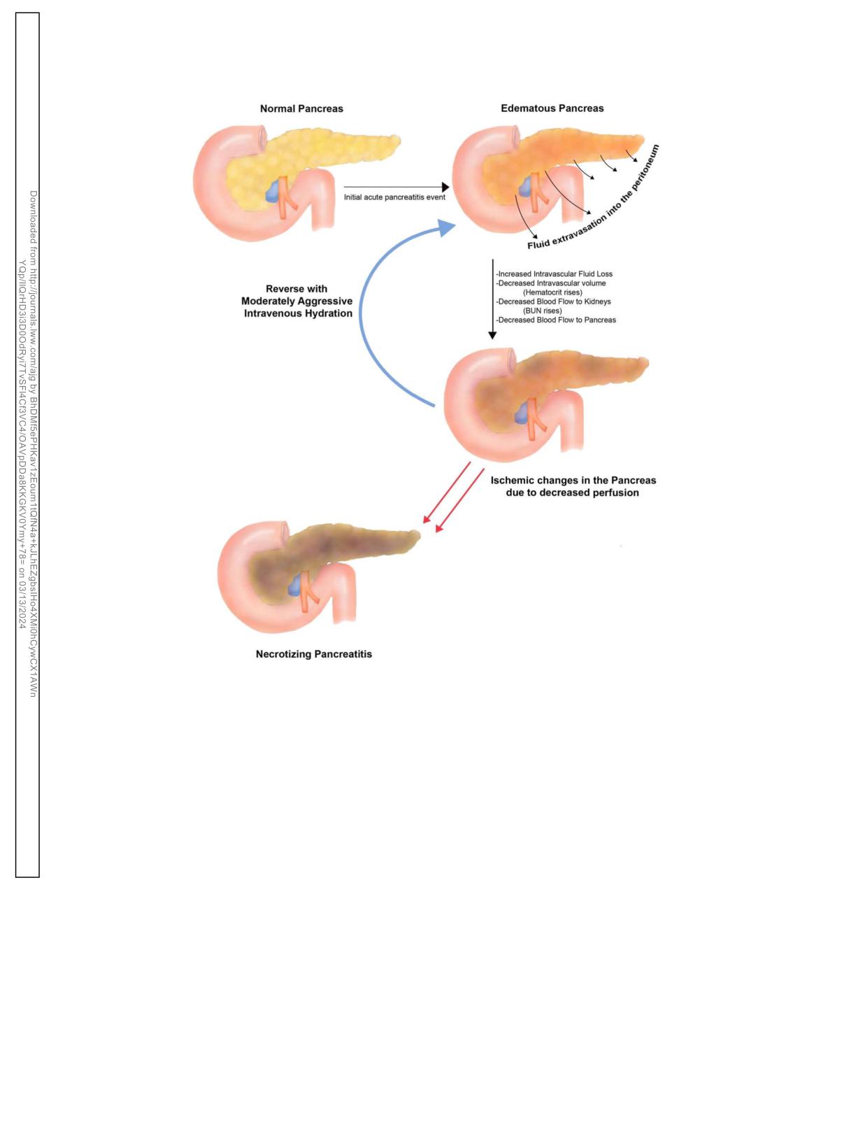
creatic necrosis (Figure 1).
specificity for severe disease (41%). Clinicians need to recognize
While there is no marker for decreasing pancreatic perfusion,
that the presence at admission or early development of SIRS in a
the rise in BUN reflects decreased renal perfusion. This can be
patient with AP warrants aggressive hydration, support, and
interpreted as a marker for decreased pancreatic perfusion. In
monitoring. For this reason, such patients should be admitted to a
addition, as the intravascular fluid leaks to the peritoneum, the
monitored bed or, if organ failure is already present, the ICU as
HCT rises as hemoconcentration develops. Early intravenous
the outcome appears improved (103).
resuscitation is essential in correcting hypovolemia, supporting
the macrocirculation and microcirculation of the pancreas to
INITIAL MANAGEMENT
prevent serious complications such as pancreatic necrosis (114).
Recommendations
On an initial review of clinical trials, conflicting conclusions
may be found regarding the benefit of early aggressive in-
3. We suggest moderately aggressive fluid resuscitation for patients
travenous hydration. However, profound differences in study
with AP. Additional boluses will be needed if there is evidence of
design explain the findings. The negative studies typically en-
hypovolemia (conditional recommendation, low quality of
rolled only patients with severe disease and/or well beyond the
evidence).
time where early aggressive intravenous hydration would have
4. We suggest using lactated Ringer solution over normal saline for
been effective (115-117). While these studies raise concerns about
intravenous resuscitation in AP (conditional recommendation,
the continuous use of aggressive hydration beyond 48 hours, and
low quality of evidence).
in patients with severe disease, the role of early hydration (within
the first 6-12 hours) was not addressed in these negative studies.
In general, the human studies that enrolled patients with mild
Key concepts
disease and provided early aggressive intravenous hydration
within the first 24 hours have shown a benefit, decreasing both
10. While we suggest all patients with AP receive moderately
morbidity and mortality (3,110,118,119). When a benefit was not
aggressive intravenous hydration of isotonic crystalloid, caution
appreciated, there were too few patients included (low power) in
is needed if a cardiovascular and/or renal comorbidity exists.
the study and/or there was not a significant difference in the
Patients should be monitored for volume overload.
amount of fluids provided to the 2 groups during the first 24 hours
11. Fluid resuscitation in patients with AP is likely more important
(92,120).
early in the course of the disease (within the first 24 hours).
Lactated Ringer solution is preferred to normal saline in the
12. Fluid volumes need to be reassessed at frequent intervals within
resuscitation and early aggressive hydration of patients with AP.
6 hours of presentation and for the next 24-48 hours with a goal
to decrease the BUN.
The benefit of using lactated Ringer solution in large-volume
resuscitation has been shown in other disease states, leading to
better electrolyte balance and outcomes (121,122). Khatua et al
Summary of evidence
(123) found that lactated Ringer solution early benefits in sys-
The initial treatment of AP depends on intravenous hydration.
temic inflammation are by providing calcium that binds ionically
This recommendation is based on expert opinion (10,104), lab-
with nonesterified fatty acids that are associated with severe
oratory experiments
(105,106), clinical indirect evidence
disease in AP. Lactate has also been shown to reduce pancreatic
(83,84,107-109) epidemiologic studies (79), and both retro-
injury in AP by decreasing inflammation (124). There are addi-
spective and prospective clinical trials (3,53,92,110). While there
tional theoretical benefits to using the more pH-balanced lactated
The American Journal of GASTROENTEROLOGY
VOLUME 119 | MARCH 2024 www.amjgastro.com
Management of Acute Pancreatitis
427
Figure 1. Role of moderately aggressive intravenous hydration in acute pancreatitis. Figure designed by Jasmine Saini, MD. BUN, blood urea nitrogen.
Ringer solution for fluid resuscitation compared with normal sa-
Monitoring patients with early aggressive intravenous hy-
line. Although both are isotonic crystalloid solutions, normal saline
dration depends on observation of clinical parameters such as
is more acidic with a pH of 5.5 and is associated with the de-
heart rate, blood pressure, and urine output. In general, in-
velopment of a nonanion gap hyperchloremic metabolic acidosis
travenous hydration providing for a decrease in the HCT (he-
and renal injury when large volumes are given (125). This has
modilution) and/or decreased BUN (increased renal perfusion)
relevance in AP where the process is premature trypsinogen acti-
have been shown to be associated with decreased morbidity and
vation that also requires a low pH. In addition, infusion of large
mortality (83,84). Although the precise timing of laboratory
volumes of normal saline has been associated with abdominal
testing and numbers for which the HCT and BUN should de-
discomfort in healthy volunteers. Thus, normal saline may exac-
crease have not been established, the latest evaluation should be
erbate the symptoms of abdominal pain associated with AP.
6-8 hours after admission (111). If an adjustment is to be made to
In 3 well-designed prospective randomized trials, lactated
the rate of hydration, it will need to be determined within this
Ringer solution has been shown to be more beneficial than nor-
time frame to assure the patient the benefit.
mal saline (53,92,119). Wu et al (92) found patients were less
A recent, elegant-designed, randomized prospective study by
likely to develop SIRS, a predictor of severe disease in patients
de-Madiera et al (116) has shown that moderate intravenous hy-
treated with lactated Ringer solution compared with those treated
dration the first 24-48 hours may be equally effective as aggressive
with normal saline. Lee et al (53) showed that patients who were
hydration. In this study, moderate hydration was less likely to cause
given lactated Ringer solution were less likely to be admitted to
volume overload when compared with early aggressive intravenous
the critical care unit and had a shorter hospitalization compared
hydration. From this study, we can conclude that in patients with
with patients with AP given normal saline. In patients who are in
no evidence of hypovolemia, an initial resuscitation rate of no more
the emergency department for a long period and inadequately
than 1.5 mL/kg of body weight per hour should be administered.
treated with early aggressive hydration, the benefit may not exist
However, in patients with hypovolemia, clinicians should admin-
and may be harmful when transferred to the floor or ICU (126).
ister a bolus of 10 mL/kg (71). While the presence of hypovolemia
© 2024 by The American College of Gastroenterology
The American Journal of GASTROENTEROLOGY
428
Tenner et al
might demand higher amounts and rates of hydration, most pa-
Summary of evidence
tients with AP will likely benefit from 3-4 L the first 24 hours,
The role of ERCP. The pathophysiology of gallstone pancreatitis
depending on body mass index. Close observation is ultimately the
involves the obstruction of the pancreatic duct by a gallstone that
key in managing patients with AP early in the course of the disease.
passes from the bile duct into the common channel as it opens
It is important to recognize that certain groups of patients, such as
into the duodenum. A persistent CBD stone (choledocholithiasis)
the older individuals and those with a history of cardiac and/or renal
can lead to persistent pancreatic duct and/or biliary tree ob-
disease, will need caution when applying hydration. Close moni-
struction, leading to necrosis and/or cholangitis (129). Although
toring for reported complications such as volume overload, pul-
intuitively, removal of obstructing gallstones from the biliary tree
monary edema, and abdominal compartment syndrome is needed
in patients with AP should reduce the complications, most gall-
(126,127). Use of central venous pressure measurement through a
stones readily pass to the duodenum and are lost in the stool
centrally placed catheter is commonly used to determine volume
(130). Most patients with gallstone pancreatitis will not benefit
status in this clinical setting. However, recent data indicate that the
from ERCP, including early ERCP.
intrathoracic blood volume index may have a better correlation with
Schepers et al (131) performed a multicenter trial to determine
cardiac index than central venous pressure, allowing more accurate
whether patients with gallstone pancreatitis and predicted severe
assessment of volume status for patients managed in the ICU.
AP (APACHE .8, Imrie .3, or C-reactive protein .150 mg/dL)
Once a patient has severe disease, there seems to be no benefit of
would benefit from early (within 24 hours) ERCP. Early ERCP
early aggressive hydration (115). Intravenous hydration in patients
was not found to decrease complications, including mortality in
with AP has been shown to be most effective early in the course of the
these patients. Yet, patients who underwent urgent ERCP were
disease (110). When severe disease develops and/or after 24 hours,
less likely to be readmitted for subsequent AP or cholangitis. The
aggressive hydration may actually be harmful (111,116,126,128).
authors concluded that urgent ERCP is indicated in this situation
While other experts and guidelines have advocated for using a term
only for cholangitis or progressive cholestasis defined by a rising
goal-directed hydration, clinicians often miss the goal failing to
bilirubin in the setting of severe or moderately severe AP (bili-
provide adequate hydration during the initial 24 hours when the
rubin .3-5 mg/dL).
moderately aggressive intravenous hydration is most important
(10,110). Keeping in mind that most patients with AP seem to have
PREVENTING POST-ERCP PANCREATITIS
mild disease, clinicians often do not appreciate the need to treat AP
Recommendations
with early hydration because the patients do not appear ill, often
having normal HCT and BUN. The goal in these patients seems to
6. We recommend rectal indomethacin to prevent post-ERCP
have been met. The problem is that AP results in an early extrava-
pancreatitis (PEP) in individuals considered to be at high risk of
sation of intravascular fluid into the peritoneum averaging 2-4 L
PEP (strong recommendation, moderate quality of evidence).
over the first 48 hours (109). If early moderately aggressive in-
7. We suggest placement of a pancreatic duct stent in patients at
travenous hydration is not provided to these patients with initially
high risk for PEP who are receiving rectal indomethacin
appearing mild AP and the disease progresses, because the BUN and/
(conditional recommendation, low quality of evidence).
or HCT rise during the first 24-36 hours, the goal is missed, and the
risk of necrosis and/or organ failure increase (108,109). Rather than
goal-directed therapy, the role of intravenous hydration is better
Summary of evidence
thought of as do not miss the goal therapy, that is, do not allow the
AP remains the most common complication of ERCP. The in-
BUN and HCT to rise within the first 24-48 hours and do not let
cidence of AP varies widely 1%-30%, depending on a variety of
SIRS and/or renal insufficiency to develop. Because once these de-
factors, including patient demographics, intraendoscopy proce-
velop, the goal of hydration was missed, and mild disease may be
dures performed, and whether the patient has received pro-
progressing to severe disease.
phylaxis (132-134). Although most patients with PEP have mild
disease, some patients have severe disease and a complicated
course, including death. There has been significant interest in
ERCP IN AP
identifying interventions that can reduce PEP.
Recommendations
In general, diagnostic ERCP should be avoided in most pa-
tients and, if needed, should be performed in Centers of Excel-
5. We suggest medical therapy over early (within the first 72 hours)
lence. Clinicians must recognize that the risk of PEP is greater in
ERCP in acute biliary pancreatitis without cholangitis (conditional
the patient with a normal caliber CBD and normal bilirubin (odds
recommendation, low quality of evidence).
ratio 3.4) when compared with a patient who is jaundiced with a
dilated CBD (odds ratio 0.2) (135). In these patients, noninvasive
MRCP or less-invasive EUS should be used because these
Key concepts
methods of evaluating the CBD are as accurate and pose no risk of
pancreatitis (136).
13. In patients with AP complicated by cholangitis, early ERCP
Interventions shown to prevent PEP include the following: (i)
within the first 24 hours has been shown to decrease morbidity
guidewire cannulation compared with contrast-guided cannula-
and mortality.
tion, (ii) pancreatic duct stents in the appropriate patient, (iii) rectal
14. In the absence of cholangitis and/or jaundice, if a CBD stone is
indomethacin suppositories, and (iv) preprocedure intravenous
suspected, MRCP or EUS should be used to screen for the
hydration (137). Guidewire cannulation, in which the bile duct and
presence of CBD stones before the use of ERCP, and diagnostic
pancreatic duct are cannulated by a guidewire inserted through a
ERCP should be avoided.
catheter (e.g., a sphincterotome), has been shown to decrease the
risk of pancreatitis (138). This is likely by avoiding hydrostatic
The American Journal of GASTROENTEROLOGY
VOLUME 119 | MARCH 2024 www.amjgastro.com
Management of Acute Pancreatitis
429
injury, but other factors may be involved. Providing clarity, in a
indomethacin and periprocedural hydration should be used in all
recent systematic review involving 15 trials, avoiding cannulation
patients before ERCP (137).
with radiocontrast agents decreased the risk of AP in most trials.
Patients undergoing ERCP who are at high risk for PEP will
The use of guidewire cannulation compared with contrast-guided
likely benefit from both rectal indomethacin and a pancreatic duct
cannulation also seems to decrease the risk of severe AP and other
stent. While a large-scale multicenter RCT showed that patients
complications, including bleeding and perforation (139).
who received rectal indomethacin alone were less likely to develop
In the appropriate patients undergoing ERCP, such as those
pancreatitis following ERCP than patients who received both rectal
with an ampullary tumor undergoing snare resection and those
indomethacin in combination with a pancreatic duct stent (152), a
undergoing endoscopic sphincterotomy, the use of a pancreatic
well-designed NIH-sponsored multicenter trial recently showed
duct stent has been shown to decrease the risk of severe PEP.
the opposite results (153). In this large trial conducted at 20 centers
Prophylactic pancreatic duct stenting is a cost-effective strategy
in the USA and Canada, 1950 patients at high risk for PEP were
for the prevention of PEP for high-risk patients (140); higher
randomly assigned to receive rectal indomethacin alone or in
incidence of severe pancreatitis has been reported in patients with
combination with a pancreatic duct stent. Patients at high risk were
failed pancreatic duct stenting (141). Yet, it is recognized that
less likely to have PEP when provided both rectal indomethacin
pancreatic duct stenting is not always technically feasible with
and a pancreatic duct stent. Therefore, prophylactic pancreatic
reported failure rate ranging from 4% to 10% (141). In addition,
duct stent placement is generally recommended in addition to
these studies supporting stent placement were unblinded and
rectal indomethacin in select patients at high risk for PEP. How-
performed by highly skilled therapeutic endoscopist, thus in-
ever, recognizing that this study was performed at tertiary care
troducing bias in favor of stenting into the results. Of more im-
centers of expertise, clinicians need to recognize the possible dif-
portance, these studies were performed before the widespread use
ficulty of placing a pancreatic duct stent in all patients at high risk
of rectal indomethacin (see further).
for PEP. A case by case approach is needed.
Multiple studies have shown that a single dose of 100 mg of
rectal indomethacin before or immediately after ERCP will pre-
THE ROLE OF ANTIBIOTICS IN AP
vent PEP in patients at high risk (134,142,143). However, in a
Recommendations
consecutive series of high-risk and low-risk patients at a single
center, no benefit to periprocedural rectal indomethacin sup-
8. We suggest against prophylactic antibiotics in patients with severe
positories was observed (144). While the benefit may not have
AP (conditional recommendation, very low quality of evidence).
been observed because of the inclusion of many patients at low
9. We suggest against fine-needle aspiration (FNA) in patients with
risk, the number needed to treat low-risk patients to prevent AP
suspected infected pancreatic necrosis (conditional
and severe AP may be still within the cost-effective range. Thus,
recommendation, very low quality of evidence).
rectal indomethacin suppositories (100 mg) should be used in all
patients undergoing ERCP, unless contraindicated (137).
Key concepts
In addition to rectal indomethacin, the use of a periprocedural
hydration with lactated Ringer solution has been shown to prevent
15. While antibiotics should not be used in patients with sterile
AP (145-147). Buxbaum (147) found that no patients developed
necrosis, antibiotics are an important part of treatment in
PEP when provided lactated Ringer solution at 3 mL/kg/hr during
infected necrosis along with debridement/necrosectomy.
the ERCP, a 20 mL/kg bolus after the procedure, followed by an 8-
16. In patients with infected necrosis, antibiotics known to penetrate
hour infusion at 3 mL/kg/hr. Similarly, 2 other randomized con-
pancreatic necrosis should be used largely to delay surgical,
trolled clinical trials showed a benefit to periprocedural intravenous
endoscopic, and radiologic drainage beyond 4 weeks. Some
hydration. Park et al (148) in a prospective randomized multicenter
patients may avoid drainage altogether because the infection
clinical trial showed that lactated Ringer solution at rate of 3 mL/mg
may completely resolve with antibiotics.
during the procedure and then 20 mL/kg bolus after the procedure
17. Routine administration of antifungal agents along with
significantly decreased the risk of PEP in average-risk to high-risk
prophylactic or therapeutic antibiotics is not needed.
patients. Similarly, Choi et al (149) found vigorous periprocedural
intravenous hydration with lactated Ringer solution reduced the
incidence and severity of PEP in average-risk and high-risk cases.
Summary of evidence
While these studies show a benefit to periprocedural infusion
Infectious complications. Infectious complications are a major
of lactated Ringer solution, the timing and additional benefit of
cause of morbidity and mortality in patients with AP, including
rectal indomethacin remains controversial. Mok et al
(142)
cholangitis (154), urinary tract infections (155), infected pseu-
conducted a randomized, double-blinded, placebo-controlled
docysts (abscesses), fluid collections (156), and infected pancre-
trial on patients at high risk of PEP, the use of a liter of intravenous
atic necrosis. SIRS that develops early in the course of AP may be
lactated Ringer solution pre-procedure with 100 mg of rectal
indistinguishable from sepsis because of fever, tachycardia,
indomethacin led to a significant decrease in postprocedure
tachypnea, and leukocytosis. When an infection is suspected,
pancreatitis. However, a larger volume of fluid and ongoing ag-
antibiotics should be given while the source of the infection is
gressive hydration post-ERCP has been shown to be not effective
being confirmed. However, once blood and other cultures are
in reducing PEP when rectal indomethacin suppositories are also
found to be negative, when no source of infection is identified,
used (150). Despite the evidence of the benefit of using rectal
antibiotics should be discontinued.
indomethacin suppositories, in a large study of more than 30,000
Sterile necrosis. The paradigm shift and controversy of using
patients, only one-third of patients were provided this method
antibiotics in AP has centered on pancreatic necrosis. When
of prophylaxis (151). When considering the costs, risks, and
compared with patients with sterile necrosis, patients with
potential benefits in light of the published literature, rectal
infected pancreatic necrosis have a higher mortality rate (mean
© 2024 by The American College of Gastroenterology
The American Journal of GASTROENTEROLOGY
430
Tenner et al
30%, range 14%-69%) (69). For this reason, preventing infection
is suspected. An immediate review of the Gram stain will often
of pancreatic necrosis is important. While some investigators
establish a diagnosis. However, it may be prudent to begin anti-
found that infection is rare in the first week after the onset of AP
biotics while awaiting microbiologic confirmation. If culture re-
(157), others have found that as many as 25% of all patients with
ports are negative, the antibiotics can be discontinued.
infected necrosis developed the infection in the first week (158).
There is some controversy as to whether a CT-FNA is necessary
Hypotension, early in the course of AP, has been believed to lead
in all patients. In many patients, the CT-FNA would not influence
to ischemia of the bowel and allow bacterial translocation from
the management of a patient (174). Many patients with sterile or
the colon leading to infection of necrosis (159). Alternatively, line
infected necrosis either improve quickly or become unstable, and
infections occurring after the first week have also been shown to
decisions on surgical intervention will not be influenced by the
lead to infection of necrosis (160).
results of the aspiration. In addition, antibiotics can be started for
Although early unblinded trials suggested a benefit in providing
suspected infection on clinical grounds even without the FNA of
antibiotics to patients with sterile necrosis by preventing infectious
the pancreatic necrosis because a negative aspiration would still
complications (155,161,162), subsequent better-designed trials have
make the antibiotic use necessary due to clinical suspicion (175). In
consistently failed to show a benefit (163-166). There have been 11
proven infection by blood or other body fluid cultures or by the
prospective randomized trials of evaluating the use of prophylactic
presence of gas in the pancreatic necrosis, the need for antibiotics is
antibiotics in severe AP, with rigorous study design, participants, and
clear. Because the infection will likely seed the necrosis, and the
outcome measures since 1993. Similarly, there were 10 meta-analyses
necrosis will be difficult to penetrate, antibiotics chosen should be
reported since 2006 describing the abovementioned RCT, although
known to penetrate the necrosis, such as carbapenems, quinolones,
the number of RCT in each meta-analysis varied depending on the
cephalosporins, and metronidazole (67,155,160,161). Routine
year of publication of meta-analysis and the selection criteria used for
administration of antifungal agents along with prophylactic or
choosing the RCT in each meta-analysis. Of interest, earlier meta-
therapeutic antibiotics is also not needed.
analyses and RCT reported a benefit with prophylactic antibiotic use
in terms of mortality, infection of pancreatic necrosis, and extrap-
NUTRITION IN AP
ancreatic infections; however, all the 3 placebo-controlled, double-
Recommendations
blind RCT, 5 of the 9 meta-analyses published after 2006, and 2 of the
recent guidelines (British Society of Gastroenterology and ACG
10. In patients with mild AP, we suggest early oral feeding (within
guidelines) (104,167) did not recommend the use of prophylactic
24-48 hours) as tolerated by the patient compared with the
antibiotics because of lack of benefit in the abovementioned
traditional nothing-by-mouth approach (conditional
outcomes.
recommendation, low quality of evidence).
Infected necrosis. The role of antibiotics in patients with necro-
11. In patients with mild AP, we suggest initial oral feeding with low-
tizing AP now focuses on the presence of infection. The concept
fat solid diet rather than a stepwise liquid to solid approach
(conditional recommendation, low quality of evidence).
that infected pancreatic necrosis requires prompt surgical de-
bridement has also been challenged by multiple reports and case
series showing that antibiotics alone can lead to resolution of in-
Key concepts
fection and, in select patients, avoid surgery altogether (168-170).
Pooling 11 studies that include 1,136 patients, there is a significant
18. Enteral nutrition in patients with moderately severe or severe AP
correlation between the timing of surgery and mortality. In general,
seems to prevent infectious complications.
in clinically stable patients, it seems that postponing necrosectomy
19. Parenteral nutrition should be avoided, unless the enteral route
in stable patients with antibiotics until 30 days after initial hospital
is not possible, not tolerated, or not meeting the caloric needs.
admission is associated with a decreased mortality.
20. Using a nasogastric rather than nasojejunal route for delivery of
Current consensus is that surgery should be performed on
enteral feeding is preferred because of comparable safety and
clinically unstable patients with infected necrosis. However, in
efficacy.
most patients, those clinically stable, the initial management of
infected necrosis should be a 30-day course of antibiotics before
surgery to allow the inflammatory reaction to become better or-
Summary of evidence
ganized (171). At this time, for a necrotic collection with a well-
Nutrition in mild AP. The long-held opinion that patients with AP
defined wall and liquefied material within, the decision and
should be nothing by mouth was based on the experience from other
method of drainage can be considered, including endoscopic,
acute abdominal conditions. The idea was to avoid food-induced
radiologic, and/or surgical intervention. If there is no response to
stimulation of pancreatic exocrine function, to decrease in-
such antibiotics in a short time or if the clinical situation deteri-
flammation and hasten recovery, and to place the pancreas at rest.
orates, necrosectomy/debridement should be performed. The
The historical practice was to wait until pain is minimal and enzymes
concept that urgent surgery is required in all patients found to
normalize or trend downward before oral feeding can be started. Oral
have infected necrosis is no longer valid.
feeding was gradually increased from clear liquid diet to soft and then
The role of CT-guided FNA. The technique of CT-guided FNA
to low-fat solid diet before discharge. It has been subsequently rec-
(CT-FNA) has proven to be safe, effective, and accurate in dis-
ognized that oral feeding maintains gut mucosal integrity and pre-
tinguishing infected and sterile necrosis (172,173). Because patients
vents translocation of bacteria from the gut lumen into the inflamed/
with infected necrosis and sterile necrosis may appear similar with
necrosed pancreatic tissue, predisposing to the serious complication
leukocytosis and fever and organ failure (67,68) it is impossible to
of infected pancreatic necrosis. This led to the concept of gut rousing
separate these entities without CT-FNA. Because the role of anti-
as opposed to gut resting (176).
biotics is best established in clinically proven infection, CT-FNA
Interest developed in early oral feeding (immediate or within
should be considered when pancreatic or extrapancreatic infection
24, 48, or 72 hours after admission) without waiting for the pain
The American Journal of GASTROENTEROLOGY
VOLUME 119 | MARCH 2024 www.amjgastro.com
Management of Acute Pancreatitis
431
and pancreatic enzymes to normalize (177-183). While most of
important in patients with AP, especially in the intensive care
these studies were conducted in patients when the treating team
setting. Nasojejunal tube placement requires interventional ra-
allowed the patients to start oral feeding, some studies applied a
diology or endoscopy and thus can be expensive. For these rea-
novel approach of starting the feeds based on hunger experienced
sons, nasogastric tube feeding maybe preferred (195).
by patients. The results of this approach seem identical
The timing of initiating enteral feeding in patients with severe
(181,184,185). For such early feeding, it is important to have
disease is controversial. While studies initially suggested a benefit
bowel sounds present and no significant nausea, vomiting, or
in preventing infectious complications, more recent studies
ileus. While most of these studies were performed in cases with
suggest that early (within the first 24 hours) initiation of enteral
mild AP, there were some studies performed in both cases with
feeding is not beneficial. Bakker et al performed a large ran-
moderately severe and severe types of disease showing a benefit to
domized trial in patients with predicted severe AP (196) and
early oral feeding (181,185,186).
found that early enteral tube feeding within 24 hours did not
Systematic reviews and meta-analyses of RCT have highlighted
reduce the rate of infection (25% vs 26%) when compared with
the benefit of early oral feeding in patients with mild, moderately
on-demand feeding. In addition, early enteral tube feeding did
severe, and severe types of AP, without the need to advance the diet
not reduce mortality (11% vs 7%).
slowly from clear liquids to solids (187,188). The universal finding
in these studies demonstrate the safety of initiating early oral
THE ROLE OF SURGERY IN AP
feeding in mild and moderately severe AP without any increase in
important clinical outcomes, such as the development of necrosis,
Key concepts
organ failure, and/or other local complications. Such an approach
is beneficial by reducing the time to initiate solid feedings, thus
21. Patients with mild acute biliary pancreatitis should undergo
reducing the hospital stay and costs. In mild AP, oral intake should,
cholecystectomy early, preferably before discharge.
in general, be restored quickly. A low-fat solid diet has been shown
22. Minimally invasive methods are preferred to open surgery for
to be safe compared with clear liquids, providing more calories
debridement and necrosectomy in stable patients with
(178). Similarly, in other randomized trials, oral feeding with a soft
symptomatic pancreatic necrosis.
diet has been found to be safe compared with clear liquids and
23. We suggest delaying any intervention (surgical, radiological,
shorten the hospital stay (189,190). A desire for food, simple
and/or endoscopic) in stable patients with pancreatic necrosis,
preferably 4 weeks, to allow for the wall of collection to mature.
hunger, can help guide clinicians’ decision when to start feedings
(185). Based on these studies, oral feedings introduced in patients
with mild AP do not need to begin with clear liquids and increase in
a stepwise manner but may begin as a low-residue, low-fat, soft diet.
Summary of evidence
However, clinicians should be aware that discharging a patient with
Cholecystectomy. In patients with mild gallstone pancreatitis,
persistent nausea despite early eating can result in readmission for
same-admission cholecystectomy has been shown to decrease
recurrent AP (191).
recurrent gallstone-related complications, with a very low risk of
Nutrition in those with moderately severe and severe AP. There
cholecystectomy-related complications (198). When evaluating
is compelling data that patients with sepsis, in general, benefit from
the literature, including 8 cohort studies and 1 randomized trial
early refeeding (192). In general, parenteral nutrition should be
describing 998 patients who were discharged rather than undergo
avoided. There have been multiple randomized trials showing that
cholecystectomy compared with early cholecystectomy, 95 (18%)
TPN is associated with infectious and other line-related compli-
were readmitted for recurrent biliary events (18% vs 0%, P ,
cations (69). Because enteral feeding maintains and prevents dis-
0.0001), including recurrent biliary pancreatitis (n 5 43, 8%)
ruption of the gut mucosal barrier, prevents disruption, and
(197). Many of these patients experienced severe disease. In ad-
prevents the translocation of bacteria that seed pancreatic necrosis,
dition to a benefit in morbidity, same-admission cholecystectomy
enteral nutrition should be begun in patients with severe AP, es-
results in substantial cost savings to the health care system (199).
pecially pancreatic necrosis (175,193). A meta-analysis of 8 ran-
Patients with pancreatic necrosis complicating biliary pan-
domized controlled clinical trials involving 381 patients found a
creatitis will require complex decision-making between the sur-
decrease in infectious complications, organ failure, and mortality in
geon and gastroenterologist. In these patients, cholecystectomy is
patients with severe AP provided enteral nutrition compared with
typically delayed to a later course in the typically prolonged
those given TPN (193). If enteral nutrition is administered by tube
hospitalization, as part of the management of the pancreatic ne-
feeds, continuous infusion is preferred over cyclic or bolus ad-
crosis if present and/or to a later date after discharge (200).
ministration (192). In addition, a small peptide-based medium-
In most patients with gallstone pancreatitis, the CBD stone
chain TG oil formula may improve tolerance (193).
passes to the duodenum. Routine ERCP is not appropriate unless
Although the use of a nasojejunal route was preferred to avoid
there is a high suspicion of a persistent CBD stone, manifested by
the gastric phase of stimulation, nasogastric enteral nutrition
an elevation in the bilirubin (201,202). Patients with mild AP,
seems safe. A systematic review describing 92 patients from 4
whose bilirubin is normal, can undergo laparoscopic cholecys-
studies on nasogastric tube feeding found that nasogastric feeding
tectomy with intraoperative cholangiography, and any remaining
was safe and well tolerated in patients with predicted severe AP
bile duct stones can be dealt with by postoperative or intra-
(194). There have been some reports of a slight increase in the risk
operative ERCP. In patients with low to moderate risk, MRCP can
of aspiration with nasogastric feeding. These patients should be
be used preoperatively; however, routine use of MRCP is un-
placed in a more upright position and be placed on aspiration
necessary. In patients with mild AP who cannot undergo surgery,
precautions. Evaluating for residuals, retained volume in the
such as older individuals and/or those with severe comorbid
stomach, is not likely to be helpful. Compared with nasojejunal
disease, biliary sphincterotomy has been shown to be effective to
feeding, nasogastric tube placement is far easier, which is
prevent recurrent biliary AP (69).
© 2024 by The American College of Gastroenterology
The American Journal of GASTROENTEROLOGY
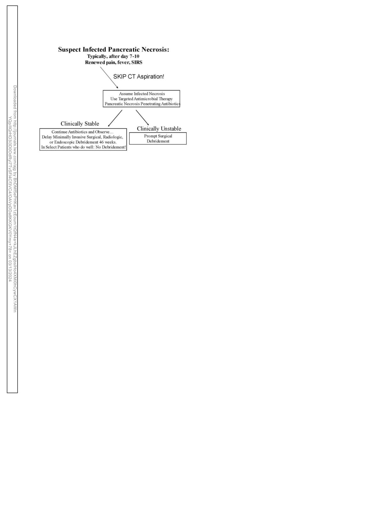
432
Tenner et al
laparoscopic surgery, radiologic catheter drainage, and endoscopy
are increasingly becoming the more common approaches. Although
these guidelines cannot discuss in detail the methods of debridement
nor the comparative effectiveness of each, due to limitations in data
and focus of this review, several generalizations are important.
In general, regardless of the method, minimally invasive ap-
proaches require the pancreatic necrosis to become better orga-
nized (171,204,206,207). Whereas early in the course of the
disease (within the first 7-10 days), pancreatic necrosis is a diffuse
solid and/or semisolid inflammatory mass, after 4 weeks, a fibrous
wall develops around the necrosis, which makes removal more
amenable to surgery, laparoscopic surgery, radiologic catheter
drainage, and/or endoscopic drainage.
Sometimes, these modalities can be combined. A well-
designed study from the Netherlands using a step-up approach
(percutaneous catheter drainage followed by video-assisted ret-
Figure 2. Late management of patients with AP. AP, acute pancreatitis; CT,
roperitoneal debridement) demonstrated the superiority of the
computed tomography; SIRS, systemic inflammatory response syndrome.
step-up approach by way of lower morbidity (less multiple organ
failure and surgical complications) and lower costs (207). The
Debridement of necrosis. Historically, open necrosectomy/
investigators confirmed a higher mortality with open surgery
debridement was the choice of treatment for infected necrosis
both as an emergency (78%) and planned (30%) compared with a
and symptomatic sterile necrosis. Decades ago, patients with
minimally invasive approach.
sterile necrosis underwent early debridement resulting in in-
Percutaneous drainage without necrosectomy may be the most
creased mortality. For this reason, early open debridement for
frequent minimally invasive method (208). The overall success
sterile necrosis was abandoned (87). For patients with infected
seems to be approximately 50% in avoiding surgery. Endoscopic
necrosis, it was falsely believed that mortality of infected necrosis
drainage of necrotic collections and later direct endoscopic
was nearly 100% if debridement was not performed urgently
necrosectomy have been reported in several large series. Two recent
(69,203). In a retrospective review of 53 patients where the me-
large multicenter studies (German and American) described the
dian time to surgery was 28 days, when necrosectomy for infected
results of direct endoscopic necrosectomy. In this endoscopic ap-
necrosis was delayed, mortality decreased 22% (157). After
proach, where endoscope is introduced into the necrotic cavity
reviewing 11 studies that included 1,136 patients, the authors also
typically through the gastric wall and necrotic tissue is removed
found a significant correlation between the timing of surgery and
under direct vision, results have been comparable (209,210). In a
mortality. It seems that postponing necrosectomy in stable pa-
recent well-designed randomized controlled clinical trial, endoscopic
tients with antibiotics until 30 days after initial hospital admission
necrosectomy seems to be superior to surgical necrosectomy (211).
is associated with a decreased mortality (168).
Regardless of the method, it must be remembered that many
The concept that infected pancreatic necrosis requires prompt
patients with sterile necrosis, and select patients with infected
surgical debridement has also been challenged by multiple reports
necrosis, seem to improve and remain asymptomatic, and no
and case series showing that antibiotics alone can lead to resolution
intervention may be necessary (212). The management of pa-
of infection and, in select patients, avoid surgery altogether
tients with necrosis is therefore very individualized, requiring
(204,205). In one report (170), of 28 patients given antibiotics for
consideration of both the clinical appearance of patients and the
the management of infected pancreatic necrosis, 16 patients avoi-
expertise available at the institution. Referral to centers of ex-
ded surgery. There were 2 deaths in the patients who underwent
pertise is of paramount importance because delaying intervention
surgery and 2 deaths in the patients who were treated with anti-
with maximal supportive care and using a minimally invasive
biotics alone. Thus, in this report, more than half the patients were
approach have both shown to be of benefit in reducing morbidity
successfully treated with antibiotics, and the mortality rates in both
and mortality in patients with acute necrotizing pancreatitis.
the surgical and nonsurgical groups were similar.
Current consensus is that the initial management of infected
ACKNOWLEDGEMENTS
necrosis for patients who are clinically stable should be a 2- to 4-
This guideline was produced in collaboration with the Practice
week course of antibiotics before surgery to allow the in-
Parameters Committee of the American College of Gastroenterology.
flammatory reaction to become better organized (171). At this
We thank our librarian Jen de Richemond for assistance in our
time, in a collection with a well-defined wall and liquefied ma-
literature search. We also give a special thanks to Jasmine Saini, MD,
terial within, the decision and method of drainage can be con-
who assisted in the research and design of the figures and collection of
sidered. For patients with symptomatic walled off pancreatic
many of the manuscripts reviewed.
necrosis, a combined multimodality approach bringing together
both minimally invasive surgery with endoscopic drainage seems
to be more effective, safer and results in a shorter hospitalization
CONFLICTS OF INTEREST
(168,203,204). Although further study is needed, the concept that
Guarantor of the article: Scott Tenner, MD, MPH, JD, FACG.
urgent surgery is required in patients found to have infected
Specific author contributions: All authors contributed to the
necrosis is no longer valid (Figure 2).
planning, data analysis, writing, and final revision of the manuscript.
Minimally invasive management of pancreatic necrosis. Mini-
Financial support: None to report.
mally invasive approaches to pancreatic necrosectomy including
Potential competing interests: None to report.
The American Journal of GASTROENTEROLOGY
VOLUME 119 | MARCH 2024 www.amjgastro.com
Management of Acute Pancreatitis
433
REFERENCES
27. Yadav D, O’Connell M, Papachristou GI. Natural history following the first
1.
Peery AF, Crockett SD, Murphy CC, et al. Burden and cost of
attack of acute pancreatitis. Am J Gastroenterol 2012;107(7):1096-103.
gastrointestinal, liver, and pancreatic diseases in the United States:
28. McAlister VC, Davenport E, Renouf E. Cholecystectomy deferral in
Update 2018. Gastroenterology 2019;156(1):254-72.e11.
patients with endoscopic sphincterotomy. Cochrane Database Syst Rev
2.
Xiao AY, Tan MLY, Wu LM, et al. Global incidence and mortality of
2007;2007:CD006233.
pancreatic diseases: A systematic review, meta-analysis, and
29. Ammann RW. The natural history of alcoholic chronic pancreatitis.
metaregression of population-based cohort studies. Lancet
Intern Med 2001;40(5):368-75.
Gastroenterol Hepatol 2016;1:45-55.
30. Aghdassi AA, Weiss FU, Mayerle J, et al. Genetic susceptibility factors
3.
Wall I, Badalov N, Baradarian R, et al. Decreased mortality in acute
for alcohol induced chronic pancreatitis. Pancreatology 2015;15(4
pancreatitis related to early aggressive hydration. Pancreas 2011;40(4):
Suppl):S23-31.
547-50.
31. Whitcomb DC. Genetic polymorphisms in alcoholic pancreatitis. Dig
4.
Guyatt GH, Oxman AD, Vist GE, et al. GRADE: An emerging consensus
Dis Sci 2005;23(3-4):247-54.
on rating quality of evidence and strength of recommendations. BMJ
32. Badalov N, Baradarian R, Iswara K, et al. Drug induced AP: An evidence
2008;336(7650):924-6.
based approach. Clin Gastroenterol Hepatol 2007;101:454-76.
5.
Banks PA, Bollen TL, Dervenis C, et al. Classification of acute
33. Parenti DM, Steinberg W, Kang P. Infectious causes of acute
pancreatitis-2012: Revision of the Atlanta classification and definitions
pancreatitis. Pancreas 1996;13(4):356-71.
by international consensus. Gut 2013;62(1):102-11.
34. Saini J, Marino D, Badalov N, et al. Drug-induced acute pancreatitis: An
6.
Rompianesi G, Hann A, Komolafe O, et al. Serum amylase and lipase and
evidence-based classification (revised). Clin Transl Gastroenterol 2023;
urinary trypsinogen and amylase for the diagnosis of AP. Cochrane
14(8):e00621.
Database Syst Rev 2017;4:CD012010.
35. Yang AL, McNabb-Baltar J. Hypertriglyceridemia and acute
7.
Clavien PA, Robert J, Meyer P, et al. Acute pancreatitis and
pancreatitis. Pancreatology 2020;20(5):795-800.
normoamylasemia. Not an uncommon combination. Ann Surg 1989;
36. Farmer RG, Winkelman EI, Brown HB, et al. Hyperlipoproteinemia and
210(5):614-20.
pancreatitis. Am J Med 1973;54(2):161-5.
8.
Winslet M, Hall C, London NJM, et al. Relation of diagnostic serum
37. Toskes PP. Hyperlipidemic pancreatitis. Gastroenterol Clin North Am
amylase levels to aetiology and severity of acute pancreatitis. Gut 1992;
1990;19(4):783-91.
33(7):982-6.
38. Murphy MJ, Sheng X, MacDonald TM, et al. Hypertriglyceridemia and
9.
Muniraj T, Dang S, Pitchumoni CS. Pancreatitis or not? Elevated lipase
acute pancreatitis. JAMA Intern Med 2013;173(2):162-4.
and amylase in ICU patients. J Crit Care 2015;30(6):1370-5.
39. Yadav D, Pitchumoni CS. Issues in hyperlipidemic pancreatitis. J Clin
10.
Crockett SD, Wani S, Gardner TB, et al. American Gastroenterological
Gastroenterol 2003;36(1):54-62.
Association Institute guideline on initial management of acute
40. Simpson WF, Adams DB, Metcalf JS, et al. Nonfunctioning pancreatic
pancreatitis. Gastroenterology 2018;154(4):1096-101.
neuroendocrine tumors presenting as pancreatitis: Report of four cases.
11.
Steinberg WM, DeVries JH, Wadden T, et al. Tu1502 longitudinal
Pancreas 1988;3(2):223-31.
monitoring of lipase and amylase in adults with type 2 diabetes and
41. Kohler H, Lankisch PG. Acute pancreatitis and hyperamylasaemia in
obesity: Evidence from two phase 3 randomized clinical trials with the
pancreatic carcinoma. Pancreas 1987;2(1):117-9.
once-daily GLP-1 analog liraglutide. Gastroenterology 2012;142(5):S-
42. Robertson JF, Imrie CW. Acute pancreatitis associated with carcinoma
850-1.
of the ampulla of Vater. Br J Surg 1987;74(5):395-7.
12.
Shah AM, Eddi R, Kothari ST, et al. Acute pancreatitis with normal
43. Cote GA, Xu H, Easler JJ, et al. Informative patterns of health-care
serum lipase: A case series. JOP 2010;11(4):369-72.
utilization prior to the diagnosis of pancreatic ductal adenocarcinoma.
13.
Kiriyama S, Gabata T, Takada T, et al. New diagnostic criteria of acute
Am J Epidemiol 2017;186(8):944-51.
pancreatitis. J Hepatobiliary Pancreat Sci 2010;17(1):24-36.
44. Alhobayb T, Peravali R, Ashkar M. The relationship between acute and
14.
Lippi G, Valentino M, Cervellin G. Laboratory diagnosis of acute
chronic pancreatitis with pancreatic adenocarcinoma: Review. Diseases
pancreatitis: In search of the Holy Grail. Crit Rev Clin Lab Sci 2012;
2021;9(4):93.
49(1):18-31.
45. Tandon M, Topazian M. Endoscopic ultrasound in idiopathic acute
15.
Reisman A, Cho HJ, Holzer H. Unnecessary repeat enzyme testing in acute
pancreatitis. Am J Gastroenterol 2001;96(3):705-9.
pancreatitis: A teachable moment. JAMA Intern Med 2018;178(5):702-3.
46. Guda NM, Muddana V, Whitcomb DC, et al. Recurrent acute
16.
Balthazar EJ. Acute pancreatitis: Assessment of severity with clinical and
pancreatitis: International state-of-the-science conference with
CT evaluation. Radiology 2002;223(3):603-13.
recommendations. Pancreas 2018;47(6):653-66.
17.
Arvanitakis M, Delhaye M, Bali M, et al. The role of magnetic resonance
47. Hallensleben N, Umans D, Bouwense SA, et al; Dutch Pancreatitis Study
imaging in the assessment of acute pancreatitis: Correlation with
Group. The diagnostic work-up and outcomes of ‘presumed’ idiopathic
computed tomography and with clinical severity and outcome.
acute pancreatitis: A post-hoc analysis of a multicentre observational
Gastroenterology 2003;124(4):A83-A84.
cohort. United European Gastroenterol J 2020;8(3):340-50.
18.
Zaheer A, Singh VK, Qureshi RO, et al. The revised Atlanta classification
48. Umans DS, Rangkuti CK, Sperna Weiland CJ, et al. Endoscopic
for acute pancreatitis: Updates in imaging terminology and guidelines.
ultrasonography can detect a cause in the majority of patients with
Abdom Imaging 2012;38(1):125-36.
idiopathic acute pancreatitis: A systematic review and meta-analysis.
19.
Bollen TL, Singh VK, Maurer R, et al. Comparative evaluation of the
Endoscopy 2020;52(11):955-64.
modified CT severity index and CT severity index in assessing severity of
49. Yusoff IF, Raymond G, Sahai AV. A prospective comparison of the yield
acute pancreatitis. AJR Am J Roentgenol 2011;197(2):386-92.
of EUS in primary vs. recurrent idiopathic acute pancreatitis.
20.
Stimac D, Miletic D, Radic M, et al. The role of nonenhanced magnetic
Gastrointest Endosc 2004;60(5):673-8.
resonance imaging in the early assessment of acute pancreatitis. Am J
50. Wan J, Ouyang Y, Yu C, et al. Comparison of EUS with MRCP in
Gastroenterol 2007;102(5):997-1004.
idiopathic acute pancreatitis: A systematic review and meta-analysis.
21.
Lankisch PG, Assmus C, Lehnick D, et al. AP: Does gender matter? Dig
Gastrointest Endosc 2018;87(5):1180-8.e9.
Dis Sci 2001;46(11):2470-4.
51. Magnusdottir BA, Baldursdottir MB, Kalaitzakis E, et al. Risk factors for
22.
Gullo L, Migliori M, Olah A, et al. Acute pancreatitis in five European
chronic and recurrent pancreatitis after first attack of acute pancreatitis.
countries: Etiology and mortality. Pancreas 2002;24(3):223-7.
Scand J Gastroenterol 2019;54(1):87-94.
23.
Yadav D, Lowenfels AB. Trends in the epidemiology of the first attack of
52. Stigliano S, Belisario F, Piciucchi M, et al. Recurrent biliary AP is
acute pancreatitis: A systematic review. Pancreas 2006;33(4):323-30.
frequent in a real-world setting. Dig Liver Dis 2018;50:77-82.
24.
Gloor B, Müller CA, Worni M, et al. Late mortality in patients with
53. Lee A, Ko C, Buitrago C, et al. Lactated ringers vs normal saline
severe acute pancreatitis. Br J Surg 2002;88(7):975-9.
resuscitation for mild acute pancreatitis: A randomized trial.
25.
Signoretti M, Baccini F, Piciucchi M, et al. Repeated transabdominal
Gastroenterology 2021;160(3):955-7.e4.
ultrasonography is a simple and accurate strategy to diagnose a biliary
54. Stevens CL, Abbas SM, Watters DA. How does cholecystectomy influence
etiology of acute pancreatitis. Pancreas 2014;43(7):1106-10.
recurrence of idiopathic AP? J Gastrointest Surg 2016;12:1997-2001.
26.
Moreau JA, Zinsmeister AR, Melton LJ, et al Gallstone pancreatitis and
55. Garg PK, Tandon RK, Madan K, et al. Is biliary microlithiasis a
the effect of cholecystectomy: A population-based cohort study. Mayo
significant cause of idiopathic recurrent AP? A long-term follow-up
Clin Proc 1988;63(5):466-73.
study. Clin Gastroenterol Hepatol 2007;5(1):75-9.
© 2024 by The American College of Gastroenterology
The American Journal of GASTROENTEROLOGY
434
Tenner et al
56.
Räty S, Pulkkinen J, Nordback I, et al. Can laparoscopic cholecystectomy
pancreatitis in a European cohort. J Gastroenterol Hepatol 2017;32(9):
prevent recurrent idiopathic AP? A prospective randomized multicenter
1649-56.
trial. Ann Surg 2015;262(5):736-41.
82.
Rasch S, Pichlmeier EM, Phillip V, et al. Prediction of outcome in acute
57.
Trna J, Vege SS, Pribramska V, et al. Lack of significant liver enzyme
pancreatitis by the qSOFA and the new ERAP score. Dig Dis Sci 2022;
elevation and gallstones and/or sludge on ultrasound on day 1 of acute
67(4):1371-8.
pancreatitis is associated with recurrence after cholecystectomy: A
83.
Brown A, Orav J, Banks PA. Hemoconcentration is an early marker for
population-based study. Surgery 2012;151(2):199-205.
organ failure and necrotizing pancreatitis. Pancreas 2000;20(4):367-72.
58.
Umans DS, Hallensleben ND, Verdonk RC, et al. Recurrence of
84.
Wu BU, Johannes RS, Sun X, et al. Early changes in blood urea nitrogen
idiopathic acute pancreatitis after cholecystectomy: Systematic review
predict mortality in acute pancreatitis. Gastroenterology 2009;137(1):
and meta-analysis. Br J Surg 2020;107(3):191-9.
129-35.
59.
Cote GA, Imperiale TF, Schmidt SE, et al. Similar efficacies of biliary,
85.
Steinberg W, Tenner S. Acute pancreatitis. N Engl J Med 1994;330(17):
with or without pancreatic, sphincterotomy in treatment of idiopathic
1198-210.
recurrent acute pancreatitis. Gastroenterology 2012;143(6):1502-9.e1.
86.
Lankisch PG, Mahlke R, Blum T, et al. Hemoconcentration: An early
60.
Steinberg WM, Chari ST, Forsmark CE, et al. Controversies in clinical
marker of severe and/or necrotizing pancreatitis? A critical appraisal.
pancreatology: Management of acute idiopathic recurrent pancreatitis.
Am J Gastroenterol 2001;96(7):2081-5.
Pancreas 2003;27(2):103-17.
87.
Frossard JL, Hadengue A, Pastor CM. New serum markers for the
61.
Badalov N, Tenner S, Baillie J. The Prevention, recognition and
detection of severe acute pancreatitis in humans. Am J Respir Crit Care
treatment of post-ERCP pancreatitis. J Pancreas 2009;10(2):88-97.
Med 2001;164(1):162-70.
62.
Cotton PB, Pauls Q, Keith J, et al. Effect of endoscopic sphincterotomy
88.
Papachristou GI, Whitcomb DC. Inflammatory markers of disease
for suspected sphincter of Oddi dysfunction on pain-related disability
severity in acute pancreatitis. Clin Lab Med 2005;25(1):17-37.
following cholecystectomy: The EPISOD randomized clinical trial.
89.
Pando E, Alberti P, Mata R, et al. Early changes in blood urea nitrogen
JAMA 2014;311(20):2101-9.
(BUN) can predict mortality in AP: Comparative study between BISAP
63.
DiMagno MJ, Dimagno EP. Pancreas divisum does not cause
score, APACHE-II, and other laboratory markers: A prospective
pancreatitis, but associates with CFTR mutations. Am J Gastroenterol
observational study. Can J Gastroenterol Hepatol 2021;1:673-82.
2012;107(2):318-20.
90.
Dancu GM, Popescu A, Sirli R, et al. The BISAP score, NLR, CRP, or
64.
Jalaly NY, Moran RA, Fargahi F, et al. An evaluation of factors associated
BUN: Which marker best predicts the outcome of AP? Medicine
with pathogenic PRSS1, SPINK1, CTFR, and/or CTRC genetic variants
(Baltimore) 2021;100(51):e28121.
in patients with idiopathic pancreatitis. Am J Gastroenterol 2017;112(8):
91.
Balthazar EJ, Robinson DL, Megibow AJ, et al. Acute pancreatitis: Value
1320-9.
of CT in establishing prognosis. Radiology 1990;174(2):331-6.
65.
Sternby H, Bolado F, Canaval-Zuleta HJ, et al. Determinants of severity
92.
Wu BU, Hwang JQ, Gardner TH, et al. Lactated Ringer’s solution
in acute pancreatitis: A nation-wide multicenter prospective cohort
reduces systemic inflammation compared with saline in patients with
study. Ann Surg 2019;270(2):348-55.
acute pancreatitis. Clin Gastroenterol Hepatol 2011;9(8):710-7.e1.
66.
Bradley EL III. A clinically based classification system for acute
93.
Funnell IC, Bornman PC, Weakley SP, et al. Obesity: An important
pancreatitis. Summary of the International Symposium on Acute
prognostic factor in acute pancreatitis. Br J Surg 1993;80(4):484-6.
Pancreatitis, Atlanta, GA, September 11 through 13, 1992. Arch Surg
94.
Heller SJ, Noordhoek E, Tenner SM, et al. Pleural effusion as a predictor
1993;128(5):586-90.
of severity in acute pancreatitis. Pancreas 1997;15(3):222-5.
67.
Tenner S. Initial management of acute pancreatitis: Critical issues during
95.
Tran DD, Cuesta MA. Evaluation of severity in patients with acute
the first 72 hours. Am J Gastroenterol 2004;99(12):2489-94.
pancreatitis. Am J Gastroenterol 1992;87(5):604-8.
68.
Perez A, Whang EE, Brooks DC, et al. Is severity of necrotizing
96.
Singh VK, Wu BU, Bollen TL, et al. Early systemic inflammatory
pancreatitis increased in extending necrosis and infected necrosis?
response syndrome is associated with severe acute pancreatitis. Clin
Pancreas 2002;25(3):229-33.
Gastroenterol Hepatol 2009;7(11):1247-51.
69.
Banks PA, Freeman ML; Practice Parameters Committee of the
97.
Mann DV, Hershman MJ, Hittinger R, et al. Multicentre audit of death
American College of Gastroenterology. Practice guidelines in acute
from acute pancreatitis. Br J Surg 1994;81(6):890-3.
pancreatitis. Am J Gastroenterol 2006;101(10):2379-400.
98.
Mutinga M, Rosenbluth A, Tenner SM, et al. Does mortality occur early
70.
Tenner S, Banks PA. Acute pancreatitis: Nonsurgical management.
or late in AP? Int J Pancreatol 2000;28(2):91-5.
World J Surg 1997;21(2):143-8.
99.
Mofidi R, Duff MD, Wigmore SJ, et al. Association between early
71.
Gardner TB. Fluid resuscitation in AP: Going over the waterfall. N Engl J
systemic inflammatory response, severity of multiorgan dysfunction and
Med 2022;387:1039-40.
death in acute pancreatitis. Br J Surg 2006;93(6):738-44.
72.
Mounzer R, Langmead CJ, Wu BU, et al. Comparison of existing clinical
100.
Buter A, Imrie CW, Carter CR, et al. Dynamic nature of early organ
scoring systems to predict persistent organ failure in patients with acute
dysfunction determines outcome in acute pancreatitis. Br J Surg 2002;
pancreatitis. Gastroenterology 2012;142(7):1476-82; quiz e15-6.
89(3):298-302.
73.
Yang CJ, Chen J, Phillips AR, et al. Predictors of severe and critical acute
101.
Johnson CD, Abu-Hilal M. Persistent organ failure during the first week
pancreatitis: A systematic review. Dig Liver Dis 2014;46(5):446-51.
as a marker of fatal outcome in acute pancreatitis. Gut 2004;53(9):
74.
Yakah W, Shah I, Skelton-Badlani D, et al. Circulating mitochondrial
1340-4.
DNA as a diagnostic biomarker for predicting disease severity in patients
102.
Lytras D, Manes K, Triantopoulou C, et al. Persistent early organ failure:
with acute pancreatitis. Gastroenterology 2023;164(6):1009-11.e3.
Defining the high risk group of patients with severe AP. Pancreas 2008;
75.
Forsmark CE, Vege SS, Wilcox CM. Acute pancreatitis. N Engl J Med
36(3):249-54.
2016;375(20):1972-81.
103.
Russell PS, Mittal A, Brown L, et al. Admission, management and
76.
Bradley EL III. Predicting clinical severity in acute pancreatitis:
outcomes of acute pancreatitis in intensive care. ANZ J Surg 2017;
Addressing the admission dilemma. Pancreas 2022;51(2):114-6.
87(12):E266-E270.
77.
Vege SS, DiMagno MJ, Forsmark CE, et al. Initial medical treatment of
104.
Tenner S, Baillie J, DeWitt J, et al; American College of Gastroenterology.
acute pancreatitis: American Gastroenterological Association Institute
American College of Gastroenterology guideline: Management of acute
technical review. Gastroenterology 2018;154(4):1103-39.
pancreatitis. Am J Gastroenterol 2013;108(9):1400-15; 1416.
78.
Dimagno MJ, Wamsteker EJ, Rizk RS, et al. A combined paging alert and
105.
Kerner T, Vollmar B, Menger MD, et al. Determinants of pancreatic
web-based instrument alters clinician behavior and shortens hospital length
microcirculation in acute pancreatitis in rats. J Surg Res 1996;62(2):
of stay in acute pancreatitis. Am J Gastroenterol 2014;109(3):306-15.
165-71.
79.
Wu BU, Johannes RS, Sun X, et al. The early prediction of mortality in
106.
Bassi D, Kollias N, Fernandez-del Castillo C, et al. Impairment of
acute pancreatitis: A large population-based study. Gut 2008;57(12):
pancreatic microcirculation correlates with the severity of acute
1698-703.
experimental pancreatitis. J Am Coll Surg 1994;179(3):257-63.
80.
Wilms H, Mittal A, Haydock MD, et al. A systematic review of goal
107.
Bize P, Platon A, Becker C, et al. Perfusion measurement in acute
directed fluid therapy: Rating of evidence for goals and monitoring
pancreatitis using dynamic perfusion MDCT. AJR Am J Roentgenol
methods. J Crit Care 2014;29(2):204-9.
2006;186(1):114-8.
81.
Valverde-López F, Matas-Cobos AM, Alegría-Motte C, et al. BISAP,
108.
Koutroumpakis E, Wu BU, Bakker OJ, et al. Admission hematocrit and
RANSON, lactate and others biomarkers in prediction of severe acute
rise in blood urea nitrogen at 24 h outperform other laboratory markers
The American Journal of GASTROENTEROLOGY
VOLUME 119 | MARCH 2024 www.amjgastro.com
Management of Acute Pancreatitis
435
in predicting persistent organ failure and pancreatic necrosis in acute
133.
Mazaki T, Mado K, Masuda H, et al. Prophylactic pancreatic stent
pancreatitis: A post hoc analysis of three large prospective databases. Am
placement and post-ERCP pancreatitis: An updated meta-analysis.
J Gastroenterol 2015;110(12):1707-16.
J Gastroenterol 2014;49(2):343-55.
109.
Sinha A, Quesada-Vazquez N, Faghih M, et al. Early predictors of fluid
134.
Elmunzer BJ, Scheiman JM, Lehman GA, et al. A randomized trial of
sequestration in acute pancreatitis: A validation study. Pancreas 2016;
rectal indomethacin to prevent post-ERCP pancreatitis. N Engl J Med
45(2):306-10.
2012;366(15):1414-22.
110.
Gardner TB, Vege SS, Chari ST, et al. Faster rate of initial fluid
135.
Mehta SN, Pavone E, Barkun JS, et al. Predictors of post-ERCP
resuscitation in severe acute pancreatitis diminishes in-hospital
complications in patients with suspected choledocholithiasis.
mortality. Pancreatology 2009;9:770-6.
Endoscopy 1998;30(5):457-63.
111.
Garg PK, Mahapatra SJ. Optimum fluid therapy in acute pancreatitis
136.
Freeman ML, Guda NM. Prevention of post-ERCP pancreatitis: A
needs an alchemist. Gastroenterology 2021;160(3):655-9.
comprehensive review. Gastrointest Endosc 2004;59(7):845-64.
112.
Dumnicka P, Maduzia D, Ceranowicz P, et al. The interplay between
137.
Buxbaum JL, Freeman M, Amateau SK, et al. American Society for
inflammation, coagulation and endothelial injury in the early phase of
Gastrointestinal Endoscopy guideline on post-ERCP pancreatitis
acute pancreatitis: Clinical implications. Int J Mol Sci 2017;18(2):354.
prevention strategies: Summary and recommendations. Gastrointest
113.
Takeda K, Mikami Y, Fukuyama S, et al. Pancreatic ischemia associated
Endosc 2023;97(2):153-62.
with vasospasm in the early phase of human acute necrotizing
138.
Lella F, Bagnolo F, Colombo E, et al. A simple way of avoiding post-
pancreatitis. Pancreas 2005;30(1):40-9.
ERCP pancreatitis. Gastrointest Endosc 2004;59(7):830-4.
114.
Gardner TB, Vege SS, Pearson RK, et al. Fluid resuscitation in acute
139.
Tse F, Liu J, Yuan Y, et al. Guidewire-assisted cannulation of the
pancreatitis. Clin Gastroenterol Hepatol 2008;6(10):1070-6.
common bile duct for the prevention of post-endoscopic retrograde
115.
Mao EQ, Fei J, Peng YB, et al. Rapid hemodilution is associated with
cholangiopancreatography (ERCP) pancreatitis. Cochrane Database
increased sepsis and mortality among patients with severe acute
Syst Rev 2022;3(3):CD009662.
pancreatitis. Chin Med J (Engl) 2010;123(13):1639-44.
140.
Das A, Singh P, Sivak MV Jr, et al. Pancreatic-stent placement for
116.
de-Madaria E, Buxbaum JL, Maisonneuve P, et al. Aggressive or
prevention of post-ERCP pancreatitis: A costeffectiveness analysis.
moderate fluid resuscitation in acute pancreatitis. N Engl J Med 2022;
Gastrointest Endosc 2007;65(7):960-8.
387(11):989-1000.
141.
Freeman ML. Pancreatic stents for prevention of postendoscopic
117.
Cuéllar-Monterrubio JE, Monreal-Robles R, González-Moreno EI, et al.
retrograde cholangiopancreatography pancreatitis. Clin Gastroenterol
Nonaggressive versus aggressive intravenous fluid therapy in acute
Hepatol 2007;5(11):1354-65.
pancreatitis with more than 24 hours from disease onset: A randomized
142.
Mok SRS, Ho HC, Shah P, et al. Lactated Ringer’s solution in
controlled trial. Pancreas 2020;49(4):579-83.
combination with rectal indomethacin for prevention of post-ERCP
118.
Buxbaum JL, Quezada M, Da B, et al. Early aggressive hydration hastens
pancreatitis and readmission: A prospective randomized, double-
clinical improvement in mild acute pancreatitis. Am J Gastroenterol
blinded, placebo-controlled trial. Gastrointest Endosc 2017;85(5):
2017;112(5):797-803.
1005-13.
119.
de-Madaria E, Herrera-Marante I, Gonzalez-Camacho V, et al. Fluid
143.
Andrade-Dávila VF, Chávez-Tostado M, Dávalos-Cobián C, et al. Rectal
resuscitation with lactated Ringer’s solution vs normal saline in AP: A
indomethacin versus placebo to reduce the incidence of pancreatitis after
triple-blind, randomized, controlled clinical trial. United European
endoscopic retrograde cholangiopancreatography: Results of a
Gastroenterol J 2018;6(1):63-71.
controlled clinical trial. BMC Gastroenterol 2015;15:85.
120.
Angsubhakorn A, Tipchaichatta K, Chirapongsathorn S, et al.
144.
Levenick JM, Gordon SR, Fadden LL, et al. Rectal indomethacin does not
Comparison of aggressive versus standard intravenous hydration for
prevent post-ERCP pancreatitis in consecutive patients.
clinical improvement among patients with mild acute pancreatitis: A
Gastroenterology 2016;150(4):911-7; quiz e19.
randomized controlled trial. Pancreatology 2021;21(7):1224-30.
145.
Sagi SV, Schmidt S, Fogel E, et al. Association of greater intravenous
121.
Khajavi MR, Etezadi F, Moharari RS, et al. Effects of normal saline vs.
volume infusion with shorter hospitalization for patients with post-
lactated Ringer’s during renal transplantation. Ren Fail 2008;30(5):535-9.
ERCP pancreatitis. J Gastroenterol Hepatol 2014;29(6):1316-20.
122.
Cho YS, Lim H, Kim SH. Comparison of lactated Ringer’s solution and
146.
DiMagno MJ, Wamsteker EJ, Maratt J, et al. Do larger periprocedural
0.9% saline in the treatment of rhabdomyolysis induced by doxylamine
fluid volumes reduce the severity of post-endoscopic retrograde
intoxication. Emerg Med J 2007;24(4):276-80.
cholangiopancreatography pancreatitis. Pancreas 2014;43(4):642-7.
123.
Khatua B, Yaron JR, El-Kurdi B, et al. Ringer’s lactate prevents early
147.
Buxbaum JI, Yan A, Yeh K, et al. Aggressive hydration with lactated
organ failure by providing extracellular calcium. J Clin Med 2020;9:263.
Ringer’s solution reduces pancreatitis after endoscopic retrograde
124.
Hoque R, Farooq A, Ghani A, et al. Lactate reduces liver and pancreatic
cholangiopancreatography. Clin Gastroenterol Hepatol 2014;12(2):
injury in Toll-like receptor and inflammasome-mediated inflammation
303-7.e1.
via GPR81-mediated suppression of innate immunity. Gastroenterology
148.
Park CH, Paik WH, Park ET, et al. Aggressive intravenous hydration
2014;146(7):1763-74.
with lactated Ringer’s solution for prevention of post-ERCP pancreatitis:
125.
Semler MW, Self WH, Wanderer JP, et al. Balanced crystalloids versus
A prospective randomized multicenter clinical trial. Endoscopy 2018;
saline in critically ill adults. N Engl J Med 2018;378(9):829-39.
50(4):378-85.
126.
Li H. The value of early aggressive hydration in patients with mild acute
149.
Choi JH, Kim HJ, Lee BU, et al. Vigorous periprocedural hydration with
pancreatitis. Chin J Emerg Med 2020;28:794-7.
lactated Ringer’s solution reduces the risk of pancreatitis after retrograde
127.
Eckerwall G, Olin H, Andersson B, et al. Fluid resuscitation and
cholangiopancreatography in hospitalized patients. Clin Gastroenterol
nutritional support during severe AP in the past: What have we learned
Hepatol 2017;15(1):86-92.e1.
and how can we do better? Clin Nutr 2006;25(3):497-504.
150.
Weiland CJS, Smeets XJNM, Kievit W, et al. Aggressive fluid hydration
128.
Gad MM, Simons-Linares CR. Is aggressive intravenous fluid
plus non-steroidal anti-inflammatory drugs versus non-steroidal anti-
resuscitation beneficial in acute pancreatitis? A meta-analysis of
inflammatory drugs alone for post-endoscopic retrograde
randomized control trials and cohort studies. World J Gastroenterol
cholangiopancreatography pancreatitis: A multicentre, open-label,
2020;26(10):1098-106.
randomized, controlled trial. Lancet Gastroenterol Hepatol 2021;6:
129.
Williams E, Beckingham I, El Sayed G, et al. Updated guideline on the
350-8.
management of common bile duct stones (CBDS). Gut 2017;66(5):765-82.
151.
Issak A, Elangovan A, Ferguson RD, et al. Underutilization of
130.
Acosta JM, Ledesma CL. Gallstone migration as a cause of acute
prophylactic rectal indomethacin and pancreatic duct stent for
pancreatitis. N Engl J Med 1974;290(9):484-7.
prevention of post-ERCP Pancreatitis. Endosc Int Open 2021;9(7):
131.
Schepers NJ, Hallensleben NDL, Besselink MG, et al. Urgent endoscopic
e979-e985.
retrograde cholangiopancreatography with sphincterotomy versus
152.
Choksi NS, Fogel EL, Cote GA, et al. The risk of post-ERCP pancreatitis
conservative treatment in predicted severe acute gallstone pancreatitis (APEC):
and the protective effect of rectal indomethacin in cases of attempted but
A multicentre randomised controlled trial. Lancet 2020;396(10245):167-76.
unsuccessful prophylactic pancreatic stent placement. Gastrointest
132.
Choudhary A, Bechtold ML, Arif M, et al. Pancreatic stents for
Endosc 2015;81(1):150-5.
prophylaxis against post-ERCP pancreatitis: A meta-analysis and
153.
Elmunzer LD, Serrano J, et al. Indomethacin with or without
systematic review. Gastrointest Endosc 2011;73(2):275-82.
prophylactic pancreatic stent placement to prevent pancreatitis after
© 2024 by The American College of Gastroenterology
The American Journal of GASTROENTEROLOGY
436
Tenner et al
ERCP: a randomized non-inferiority trial. Lancet 2024:S0140-6736.
meal in mild acute pancreatitis. Clin Gastroenterol Hepatol 2007;5(8):
[Online ahead of print January 11, 2024.] PMID: 38219767.
946-51; quiz 886.
154.
Fan ST, Lai EC, Mok FP, et al. Early treatment of acute biliary
179.
Teich N, Aghdassi A, Fischer J, et al. Optimal timing of oral refeeding in
pancreatitis by endoscopic papillotomy. N Engl J Med 1993;328(4):
mild acute pancreatitis: Results of an open randomized multicenter trial.
228-32.
Pancreas 2010;39(7):1088-92.
155.
Saino V, Kemppainem E, Puolakkainen P, et al. Early antibiotic
180.
Lariño-Noia J, Lindkvist B, Iglesias-García J, et al. Early and/or
treatment in acute necrotizing pancreatitis. Lancet 1995;346:663-7.
immediately full caloric diet versus standard refeeding in mild acute
156.
Baril NB, Ralls PW, Wren SM, et al. Does an infected peripancreatic fluid
pancreatitis: A randomized open-label trial. Pancreatology 2014;14(3):
collection or abscess mandate operation? Ann Surg 2000;231(3):361-7.
167-73.
157.
Besselink MG, Verwer TJ, Schoenmaeckers EJP, et al. Timing of surgical
181.
Zhao XL, Zhu SF, Xue GJ, et al. Early oral refeeding based on hunger in
Intervention in necrotizing pancreatitis. Arch Surg 2007;142(12):
moderate and severe acute pancreatitis: A prospective controlled,
1194-201.
randomized clinical trial. Nutrition 2015;31(1):171-5.
158.
Petrov MS, Shanbhag S, Chakraborty M, et al. Organ failure and
182.
Dong E, Chang JI, Verma D, et al. Enhanced recovery in mild acute
infection of pancreatic necrosis as determinants of mortality in patients
pancreatitis: A randomized controlled trial. Pancreas 2019;48(2):
with acute pancreatitis. Gastroenterology 2010;139(3):813-20.
176-81.
159.
Runkel NS, Moody FG, Smith GS, et al. The role of the gut in the
183.
Horibe M, Iwasaki E, Nakagawa A, et al. Efficacy and safety of immediate
development of sepsis in acute pancreatitis. J Surg Res 1991;51(1):18-23.
oral intake in patients with mild acute pancreatitis: A randomized
160.
Beger HG, Bittner R, Block S, et al. Bacterial contamination of pancreatic
controlled trial. Nutrition 2020;74:110724.
necrosis: A perspective clinical study. Gastroenterology 1986;91(2):
184.
Li J, Xue GJ, Liu YL, et al. Early oral refeeding wisdom in patients with
433-8.
mild acute pancreatitis. Pancreas 2013;42(1):88-91.
161.
Pederzoli P, Bassi C, Vesentini S, et al. A randomized multicenter clinical
185.
Rai A, Anandhi A, Sureshkumar S, et al. Hunger-based versus
trial of antibiotic prophylaxis of septic complications in acute
conventional oral feeding in moderate and severe acute pancreatitis: A
necrotizing pancreatitis with imipenem. Surg Gynecol Obstet 1993;
randomized controlled trial. Dig Dis Sci 2022;67(6):2535-42.
176(5):480-3.
186.
Ramırez-Maldonado E, López Gordo S, Pueyo EM, et al. Immediate oral
162.
Bassi C, Pederzoli P, Vesentini S, et al. Behavior of antibiotics during
refeeding in patients with mild and moderate acute pancreatitis: A
human necrotizing pancreatitis. Antimicrob Agents Chemother 1994;
multicenter, randomized controlled trial (PADI trial). Ann Surg 2021;
38(4):830-6.
274(2):255-63.
163.
Dellinger EP, Tellado JM, Soto NE, et al. Early antibiotic treatment for
187.
Vaughn VM, Shuster D, Rogers MAM, et al. Early versus delayed feeding
severe acute necrotizing pancreatitis: A randomized, double blind,
in patients with acute pancreatitis: A systematic review. Ann Intern Med
placebo controlled study. Ann Surg 2007;245(5):674-83.
2017;166(12):883-92.
164.
Jafri NS, Mahid SS, Idstein SR, et al. Antibiotic prophylaxis is not
188.
Yao Q, Liu P, Peng S, et al. Effects of immediate or early oral feeding on
protective in severe acute pancreatitis: A systematic review and meta-
acute pancreatitis: A systematic review and meta-analysis.
analysis. Am J Surg 2009;197(6):806-13.
Pancreatology 2022;22(2):175-84.
165.
Villatoro E, Bassi C, Larvin M. Antibiotic therapy for prophylaxis against
189.
Sathiaraj E, Murthy S, Mansard MJ, et al. Clinical trial: Oral feeding with
infection of pancreatic necrosis in acute pancreatitis. Cochrane Database
a soft diet compared with clear liquid diet as initial meal in mild acute
Syst Rev 2006;4:CD002941.
pancreatitis. Aliment Pharmacol Ther 2008;28(6):777-81.
166.
De Vries A, Besselink MG, Buskens E, et al. Randomized controlled trials
190.
Moraes JM, Felga GE, Chebli LA, et al. A full solid diet as the initial meal
of antibiotic prophylaxis in severe acute pancreatitis: Relationship
in mild acute pancreatitis is safe and result in a shorter length of
between methodological quality and outcome. Pancreatology 2007;
hospitalization: Results from a prospective, randomized, controlled,
7(5-6):531-8.
double-blind clinical trial. J Clin Gastroenterol 2010;44(7):517-22.
167.
UK Working Party on Acute Pancreatitis. UK guidelines for the
191.
Whitlock TL, Repas K, Tignor A, et al. Early readmission in acute
management of acute pancreatitis. Gut 2005;54(Suppl 3):iii1-9.
pancreatitis: Incidence and risk factors. Am J Gastroenterol 2010;
168.
Hartwig W, Maksan SM, Foitzik T, et al. Reduction in mortality with
105(11):2492-7.
delayed surgical therapy of severe pancreatitis. J Gastrointest Surg 2002;
192.
Doig GS, Heighes PT, Simpson F, et al. Early enteral nutrition, provided
6(3):481-7.
within 24 h of injury or intensive care unit admission, significantly
169.
Dubner H, Steinberg W, Hill M, et al. Infected pancreatic necrosis and
reduces mortality in critically ill patients: A meta-analysis of randomised
peripancreatic fluid collections: Serendipitous response to antibiotics
controlled trials. Intensive Care Med 2009;35(12):2018-27.
and medical therapy in three patients. Pancreas 1996;12(3):298-302.
193.
Yi F, Ge L, Zhao J, et al. Meta-analysis: Total parenteral nutrition versus
170.
Runzi M, Niebel W, Goebell H, et al. Severe acute pancreatitis:
total enteral nutrition in predicted severe acute pancreatitis. Intern Med
Nonsurgical treatment of infected necroses. Pancreas 2005;30(3):195-9.
2012;51(6):523-30.
171.
Vege SS, Baron TH. Management of pancreatic necrosis in severe AP.
194.
Petrov MS, Pylypchuk RD, Emelyanov NV. Systematic review:
Clin Gastroenterol Hepatol 2004;99:2489-94.
Nutritional support in acute pancreatitis. Aliment Pharmacol Ther 2008;
172.
Gerzof SG, Banks PA, Robbins AH, et al. Early diagnosis of pancreatic
28(6):704-12.
infection by computed tomography guided aspiration. Gastroenterology
195.
Singh N, Sharma B, Sharma M, et al. Evaluation of early enteral feeding
1987;93(6):1315-20.
through nasogastric and nasojejunal tube in severe acute pancreatitis: A
173.
Buchler MW, Gloor B, Müller CA, et al. Acute necrotizing pancreatitis:
noninferiority randomized controlled trial. Pancreas 2012;41(1):153-9.
Treatment strategy according to the status of infection. Ann Surg 2000;
196.
Bakker OJ, van Brunschot S, van Santvoort HC, et al. Early versus on-
232(5):619-26.
demand nasoenteric tube feeding in acute pancreatitis. N Engl J Med
174.
Pappas T. Is CT guided fine needle aspiration helpful in patients with
2014;371(21):1983-93.
infected necrosis. Am J Gastroenterol 2005;100:2371-4.
197.
Van Baal MC, Besselink MG, Bakker OJ, et al. Timing of
175.
Isenmann R, Runzi M, Kron M, et al. Prophylactic antibiotic treatment
cholecystectomy after mild biliary pancreatitis: A systematic review.
in patients with predicted severe acute pancreatitis: A placebo-
Ann Surg 2012;255(5):860-6.
controlled, double-blind trial. Gastroenterology 2004;126(4):997-1004.
198.
Da Costa DW, Boerma D, Schepers NJ, et al. Same-admission versus
176.
Petrov MS, Kukosh MV, Emelyanov NV. A randomized controlled trial
interval cholecystectomy for mild gallstone pancreatitis (PONCHO): A
of enteral versus parenteral feeding in patients with predicted severe
multicenter randomized controlled trial. Lancet 2015;26:1261-8.
acute pancreatitis shows a significant reduction in mortality and in
199.
Da Costa DW, Dijksman LM, Bouwense SA, et al. Cost-effectiveness of
infected pancreatic complications with total enteral nutrition. Dig Surg
same-admission versus interval cholecystectomy after mild gallstone
2006;23(5-6):336-45.
pancreatitis in the PONCHO trial. Br J Surg 2016;103(12):1695-703.
177.
Eckerwall G, Andersson R. Early enteral nutrition in severe acute
200.
Gurusamy KS, Nagendran M, Davidson BR. Early versus delayed
pancreatitis: A way of providing nutrients, gut barrier protection,
laparoscopic cholecystectomy for acute gallstone pancreatitis. Cochrane
immunomodulation, or all of them? Scand J Gastroenterol 2001;36(5):
Database Syst Rev 2013;9:CD010326.
449-58.
201.
Ayub K, Imada R, Slavin J. Endoscopic retrograde
178.
Jacobson BC, Vander Vliet MB, Hughes MD, et al. A prospective,
cholangiopancreatography in gallstone-associated acute pancreatitis.
randomized trial of clear liquids versus low-fat solid diet as the initial
Cochrane Database Syst Rev 2004;4:CD003630.
The American Journal of GASTROENTEROLOGY
VOLUME 119 | MARCH 2024 www.amjgastro.com
Management of Acute Pancreatitis
437
202. Mark DH, Lefevre F, Flamm CR, et al. Evidence based assessment of
209. Seifert H, Biermer M, Schmitt W, et al. Transluminal endoscopic
ERCP in the treatment of pancreatitis. Gastrointest Endosc 2002;56(6
necrosectomy after acute pancreatitis: A multicentre study with long-
Suppl):S249-54.
term follow-up (the GEPARD study). Gut 2009;58(9):1260-6.
203. Adler DG, Chari ST, Dahl TJ, et al. Conservative management of infected
210. Gardner TB, Coelho-Prabhu N, Gordon SR, et al. Direct endoscopic
necrosis complicating severe acute pancreatitis. Am J Gastroenterol
necrosectomy for the treatment of walled-off pancreatic necrosis:
2003;98(1):98-103.
Results from a multi-center US series. Gastrointest Endosc 2011;73(4):
204. Sarr M, Seewald S. Do all patients with documented infected necrosis require
718-26.
necrosectomy/drainage. Clin Gastroenterol Hepatol 2010;8(12):1000-1.
211. Bakker OJ, van Santvoort HC, van Brunschot S, et al. Endoscopic
205. Gluck M, Ross A, Irani S, et al. Endoscopic and percutaneous drainage of
transgastric vs surgical necrosectomy for infected necrotizing
symptomatic walled off pancreatic necrosis reduces hospital stay and
pancreatitis; a randomized trial. JAMA 2012;307(10):1053-61.
radiographic resources. Clin Gastroenterol Hepatol 2010;8(12):1083-8.
212. Garg PK, Sharma M, Madan K, et al. Primary conservative treatment
206. Mier J, Leon EL, Castillo A, et al. Early versus late necrosectomy in severe
results in mortality comparable to surgery in patients with infected
necrotizing pancreatitis. Am J Surg 1997;173(2):71-5.
pancreatic necrosis. Clin Gastroenterol Hepatol 2010;8(12):1089-94.e2.
207. van Santvoort HC, Bakker OJ, Bollen T, et al. A conservative and
213. Ranson JH, Pasternack BS. Statistical methods for quantifying the
minimally invasive approach to necrotizing pancreatitis improves
severity of clinical acute pancreatitis. J Surg Res 1977;22(2):79-91.
outcome. Gastroenterology 2011;141(4):1254-63.
214. Papachristou GI, Muddana V, Yadav D, et al. Comparison of BISAP,
208. van Baal MC, van Santvoort HC, Bollen TL, et al. Systematic review
Ranson’s, APACHE-II, and CTSI scores in predicting organ failure,
ofpercutaneous catheter drainage as primary treatment for necrotizing
complications, and mortality in acute pancreatitis. Am J Gastroenterol
pancreatitis. Br J Surg 2011;98(1):18-27.
2010;105(2):435-41; quiz 442.
© 2024 by The American College of Gastroenterology
The American Journal of GASTROENTEROLOGY
Gastroenterology 2024;166:521-532
CLINICAL PRACTICE UPDATES
AGA Clinical Practice Update on Diet and Nutritional Therapies in
Patients With Inflammatory Bowel Disease: Expert Review
Jana G. Hashash,1 Jaclyn Elkins,2 James D. Lewis,3 and David G. Binion4
1Division of Gastroenterology and Hepatology, Mayo Clinic, Jacksonville, Florida;2Department of Nutrition, Mayo Clinic,
Jacksonville, Florida;3Division of Gastroenterology and Hepatology, Perelman School of Medicine, University of Pennsylvania,
Philadelphia, Pennsylvania; and4Division of Gastroenterology, Hepatology, and Nutrition, University of Pittsburgh, Pittsburgh,
Pennsylvania
DESCRIPTION: Diet plays a critical role in human health, but
consistently been found to decrease the rate of flares in adults
especially for patients with inflammatory bowel disease (IBD).
with IBD. A diet low in red and processed meat may reduce
Guidance about diet for patients with IBD are often contro-
ulcerative colitis flares, but has not been found to reduce
versial and a source of uncertainty for many physicians and
relapse in Crohn’s disease. BEST PRACTICE ADVICE 2: Pa-
patients. The role of diet has been investigated as a risk factor
tients with IBD who have symptomatic intestinal strictures may
for IBD etiopathogenesis and as a therapy for active disease.
not tolerate fibrous, plant-based foods (ie, raw fruits and veg-
Dietary restrictions, along with the clinical complications of
etables) due to their texture. An emphasis on careful chewing
IBD, can result in malnutrition, an underrecognized condition
and cooking and processing of fruits and vegetables to a soft,
among this patient population. The aim of this American
less fibrinous consistency may help patients with IBD who have
Gastroenterological Association (AGA) Clinical Practice Update
concomitant intestinal strictures incorporate a wider variety of
(CPU) is to provide best practice advice statements, primarily
plant-based foods and fiber in their diets. BEST PRACTICE
to clinical gastroenterologists, covering the topics of diet and
ADVICE 3: Exclusive enteral nutrition using liquid nutrition
nutritional therapies in the management of IBD, while empha-
formulations is an effective therapy for induction of clinical
sizing identification and treatment of malnutrition in these
remission and endoscopic response in Crohn’s disease, with
patients. We provide guidance for tailored dietary approaches
stronger evidence in children than adults. Exclusive enteral
during IBD remission, active disease, and intestinal failure. A
nutrition may be considered as a steroid-sparing bridge ther-
healthy Mediterranean diet will benefit patients with IBD, but
apy for patients with Crohn’s disease. BEST PRACTICE ADVICE
may require accommodations for food texture in the setting of
4: Crohn’s disease exclusion diet, a type of partial enteral
intestinal strictures or obstructions. New data in Crohn’s dis-
nutrition therapy, may be an effective therapy for induction of
ease supports the use of enteral liquid nutrition to help induce
clinical remission and endoscopic response in mild to moderate
remission and correct malnutrition in patients heading for
Crohn’s disease of relatively short duration. BEST PRACTICE
surgery. Parenteral nutrition plays a critical role in patients
ADVICE 5: Exclusive enteral nutrition may be an effective
with IBD facing acute and/or chronic intestinal failure. Regis-
therapy in malnourished patients before undergoing elective
tered dietitians are an essential part of the interdisciplinary
surgery for Crohn’s disease to optimize nutritional status and
team approach for optimal nutrition assessment and manage-
reduce postoperative complications. BEST PRACTICE ADVICE
ment in the patient population with IBD. METHODS: This
6: In patients with IBD who have an intra-abdominal abscess
expert review was commissioned and approved by the AGA
and/or phlegmonous inflammation that limits ability to achieve
Clinical Practice Updates Committee and the AGA Governing
optimal nutrition via the digestive tract, short-term parenteral
Board to provide timely guidance on a topic of high clinical
nutrition may be used to provide bowel rest in the preoperative
importance to the AGA membership and underwent internal
phase to decrease infection and inflammation as a bridge to
peer review by the CPU Committee and external peer review
definitive surgical management and to optimize surgical out-
through standard procedures of Gastroenterology. The best
comes. BEST PRACTICE ADVICE 7: We suggest the use of
practice advice statements were drawn from reviewing existing
parenteral nutrition for high-output gastrointestinal fistula,
literature combined with expert opinion to provide practical
prolonged ileus, short bowel syndrome, and for patients with
advice on the role of diet and nutritional therapies in patients
IBD with severe malnutrition when oral and enteral nutrition
with IBD. Because this was not a systematic review, formal
has been trialed and failed or when enteral access is not
rating of the quality of evidence or strength of the presented
feasible or contraindicated. BEST PRACTICE ADVICE 8: In
considerations was not performed.
patients with IBD and short bowel syndrome, long-term
parenteral nutrition should be transitioned to customized hy-
dration management (ie, intravenous electrolyte support and/
BEST PRACTICE ADVICE STATEMENTS
or oral rehydration solutions) and oral intake whenever
possible to decrease the risk of developing long-term compli-
BEST PRACTICE ADVICE 1: Unless there is a contraindication,
cations. Treatment with glucagon-like peptide-2 agonists can
all patients with IBD should be advised to follow a Mediterra-
facilitate this transition. BEST PRACTICE ADVICE 9: All pa-
nean diet rich in a variety of fresh fruits and vegetables,
tients with IBD warrant regular screening for malnutrition by
monounsaturated fats, complex carbohydrates, and lean pro-
their provider by means of assessing signs and symptoms,
teins and low in ultraprocessed foods, added sugar, and salt for
including unintended weight loss, edema and fluid retention,
their overall health and general well-being. No diet has
and fat and muscle mass loss. When observed, more complete
522
Hashash et al
Gastroenterology Vol. 166, Iss. 3
evaluation for malnutrition by a registered dietitian is indi-
vegetables, for IBD management is a substantial revision
cated. Serum proteins are no longer recommended for the
from past instructions (Figure 1, Table 1). Prior emphasis on
identification and diagnosis of malnutrition due to their lack of
a low-residue, low-fiber diet is reasonable for patients with
specificity for nutritional status and high sensitivity to inflam-
IBD who are experiencing symptomatic disease flares and
mation. BEST PRACTICE ADVICE 10: All patients with IBD
worsening abdominal symptoms, but whenever possible,
should be monitored for vitamin D and iron deficiency. Patients
long-term IBD management should attempt to reintroduce
with extensive ileal disease or prior ileal surgery (resection or
fresh fruits, vegetables, and fiber (preferably soluble fiber).
ileal pouch) should be monitored for vitamin B12 deficiency.
Recent prospective, randomized, short-term (6-12 weeks)
BEST PRACTICE ADVICE 11: All outpatients and inpatients
studies have suggested that a Mediterranean diet and a
with complicated IBD warrant co-management with a regis-
more structured specific carbohydrate diet were equally
tered dietitian, especially those who have malnutrition, short
effective in achieving symptomatic remission and calpro-
bowel syndrome, enterocutaneous fistula, and/or are requiring
tectin response (Table 1).1 Importantly, the use of a Medi-
more complex nutrition therapies (eg, parenteral nutrition,
terranean diet may mechanistically improve the diversity of
enteral nutrition, or exclusive enteral nutrition), or those on a
the gut microbiome and metabolome and hold additional
Crohn’s disease exclusion diet. We suggest that all newly
long-term health benefits, such as reduction of cardiovas-
diagnosed patients with IBD have access to a registered dieti-
cular disease, metabolic syndrome, and cancer. An inde-
tian. BEST PRACTICE ADVICE 12: Breastfeeding is associated
with a lower risk for diagnosis of IBD during childhood. A
pendent validation of the efficacy of the Mediterranean diet
healthy, balanced, Mediterranean diet rich in a variety of fruits
for patients with IBD was performed by Chicco et al.2
and vegetables and decreased intake of ultraprocessed foods
Nutritional counseling was provided to 142 patients with
have been associated with a lower risk of developing IBD.
IBD. After 6 months, both patients with UC and patients with
CD adhering to the Mediterranean diet had lower rates of
active disease, inflammatory biomarker elevation, and
Keywords: IBD; Nutrition; Diet; CDED; Parenteral Nutrition.
improved quality of life.
To date, there is no consistent evidence supporting the
avoidance of gluten in patients with IBD in the absence of a
he central purpose of the gastrointestinal tract is
celiac disease diagnosis or suspected gluten sensitivity.
T
nutrition, and this essential function is often
Although the use of a low fermentable oligo-, di-, and
compromised in patients with inflammatory bowel disease
monosaccharide and polyols (FODMAP) diet was found to
(IBD). There is growing recognition of the role of diet in the
improve symptoms in patients with IBD in a prospective,
care of patients with IBD, as both an etiopathogenic risk factor
randomized trial, this may be accompanied by potential
and, more recently, as a disease-modifying modality. Histori-
negative long-term consequences.3 The low-FODMAP diet
cally, there was limited guidance regarding diet for patients
results in the reduction of certain fecal microbiome organ-
with IBD. Other than to counsel on avoiding foods that worsen
isms and reduced generation of the short-chain fatty acid
symptoms and to avoid foods that may predispose to
butyrate, a key nutrient for gut epithelial health. Research
obstruction in those with strictures, health care providers had
has found that the organisms that are diminished in patients
limited diet-related input to give their patients. Although such
on a low-FODMAP diet tend to be associated with endo-
dietary advice may help improve symptoms in the acute
scopic and clinical remission when found in abundance,
setting, these approaches frequently led patients with IBD to
raising concern about the long-term effects of low-FODMAP
avoid what are traditionally considered healthy foods, even
diets.4-6 Thus, short-term use of a reduced-fiber, low-FOD-
after achieving clinical remission. New insights have resulted
MAP dietary approach during a symptomatic IBD flare may
from the investigation of diet and nutrition in the overall care
be helpful, but as patients achieve symptom resolution, we
of patients with Crohn’s disease (CD) and ulcerative colitis
propose that a return to a healthy Mediterranean-style diet
(UC). The aim of this American Gastroenterological Associa-
is in the best long-term interest of patients with IBD.
tion Clinical Practice Update is to provide best practice advice
Adherence to a healthy, balanced Mediterranean diet
statements on the role of diet and nutritional therapies in the
will confer the additional benefit of effectively reducing
treatment of patients with IBD, with a focus on common
intake of ultraprocessed foods, which often contain added
clinical scenarios encountered during IBD care.
sugar, excess salt, and other food additives. High con-
Best Practice Advice 1: Unless there is a contrain-
sumption of ultraprocessed foods has been implicated in the
dication, all patients with IBD should be advised to follow
a Mediterranean diet rich in a variety of fresh fruits and
Abbreviations used in this paper: CD, Crohn’s disease; CDED, Crohn’s
vegetables, monounsaturated fats, complex carbohydrates,
disease exclusion diet; EEN, exclusive enteral nutrition; EN, enteral
nutrition; ESPEN, European Society for Parenteral and Enteral Nutrition;
and lean proteins and low in ultraprocessed foods, added
FODMAP, fermentable oligo-di-and monosaccharide and polyols; GLP-2,
sugar, and salt for their overall health and general well-
glucagon-like peptide-2; IBD, inflammatory bowel disease; IVF, intrave-
being. No diet has consistently been found to decrease
nous fluids; NPO, nil per os (nothing by mouth); PEN, partial enteral
nutrition; PN, parenteral nutrition; PO, per os (by mouth); RD, registered
the rate of flares in adults with IBD. A diet low in red and
dietitian; SBS, short bowel syndrome; UC, ulcerative colitis.
processed meat may reduce ulcerative colitis flares, but
Most current article
has not been found to reduce relapse in Crohn’s disease.
© 2024 by the AGA Institute.
The guidance for consumption of healthy eating pat-
0016-5085/$36.00
terns, such as the Mediterranean diet rich in fruits and
March 2024
AGA Clinical Practice Update on Diet and IBD
523
Figure
1. Optimal nutri-
tional approach by clinical
state of IBD.
emergence of health problems throughout the world,
from Crohn’s and Colitis Canada and Crohn’s and Colitis
including chronic inflammation, and IBD with CD has the
Foundation.11,12
strongest association,7,8 At present, it is not known which
Best Practice Advice 2: Patients with IBD who have
dietary components of the Mediterranean diet that are
symptomatic intestinal strictures may not tolerate
emphasized vs those that are minimized underlie its
fibrous, plant-based foods (ie, raw fruits and vegetables)
overall efficacy in improving the health of patients with
due to their texture. An emphasis on careful chewing and
IBD. Specific dietary components that patients with IBD
cooking and processing of fruits and vegetables to a soft,
should be cautioned to avoid are sugar-sweetened bev-
less fibrinous consistency may help patients with IBD
erages, which have been linked to etiopathogenic risk and
who have concomitant intestinal strictures incorporate a
a more severe multiyear clinical course of IBD in a recent
wider variety of plant-based foods and fiber in their diets.
prospective cohort study.9,10 IBD-specific diet and nutri-
Inflammatory injury of the gastrointestinal tract can lead
tion guidance for patients and caregivers are available to intestinal remodeling with scarring and strictures,
Table 1.Diets That Have Been Studied in Patients With Inflammatory Bowel Disease
Diet
Description/Rationale
Comments
The Mediterranean diet
Plant-focused diet emphasizing variety of whole grains, fruits, and vegetables.
A recent study (2021)1 suggested that for adults with mild to moderate CD, the
Main fat source is from fish, olive oil, nuts, and seeds.
Mediterranean diet has similar efficacy to a specific carbohydrate diet.
Lean protein sources are from low-fat dairy, poultry, fish, shellfish, beans,
The Mediterranean diet aligns with a moderate- to high-fiber diet for those in
and/or legumes.
remission.
Higher saturated fat containing meats (ie, red meat) are consumed at much
The Mediterranean diet has demonstrated health benefits separate from IBD,
lower frequency and quantity.
such as reduced cardiovascular disease incidence.
Specific carbohydrate
Nutritionally complete grain-free diet, low in sugar and lactose. Restricts all
Challenging to follow.
diet
hard-to-digest carbohydrates, only eating those that are easy to break down.
There have been limited large-scale studies showing evidence of benefit.
Examples of included foods:
Relatively similar efficacy to Mediterranean diet in DINE-CD (Diet to Induce
Additive free meat and oils (white vinegar, cider, and mustard)
Remission in Crohn’s Disease) trial.
Sugar-free coffee, tea, nut butters, and juice
Low-lactose dairy
Nonstarchy vegetables
Examples of foods not allowed:
Grains and grain products
Candy or foods made with high-fructose corn syrup
High-lactose dairy
Starchy vegetables
Sugars, excluding honey
Hypothesis is that these foods fuel “bad” bacteria in the gut, and thus avoiding
them aids “good” bacterial survival.
Low-FODMAP diet
Elimination rechallenge diet that limits fermentable oligosaccharides,
May be worth trying in patients with IBD who have concomitant IBS-like
disaccharides, monosaccharides, and polyols, which are short-chain
symptoms.
carbohydrates (sugars) that the small intestine absorbs poorly. These are
omitted from the diet for up to 8 wk, then reintroduced 1 at a time.
CDEDa
Whole foods diet designed to limit foods that may adversely affect the
May be worth attempting in patients with mild to moderate CD with short
microbiome or alter intestinal barrier function. Diet is initiated in 3 phases
duration of flares.
and each phase is 6 wk long and includes partial EN (liquid formula either
Allows for some solid foods compared with the 100% liquid nature of EEN;
by mouth or enterically infused).
may improve compliance and be easier to follow.
Phase 1:
Mandatory intake of fish, chicken breast, and eggs
Allows rice, cooled potatoes, tomatoes, onion, garlic, ginger, olive oil, and canola oil
Limited quantities of cucumber, carrots, spinach, lettuce, bananas, apples,
avocados, strawberries, melon, and citrus juices
Phase 2:
Phase 1 foods þ tuna, whole-grain bread, oats, yams, and red peppers
Certain vegetables, beans, peas, turnips, and parsnips are reintroduced after wk 10
Phase 3:
“Maintenance phase”
Phase 1 and 2 foods þ more seafood, eggs, cocoa, coffee, grains, some dairy,
and alcohol if tolerated
aPlease refer to Figure
2.
March 2024
AGA Clinical Practice Update on Diet and IBD
525
making fibrous, plant-based foods a trigger for obstructive
components of PEN regimens.24,25 The Crohn’s disease
symptoms (Figure 1). Although previous IBD dietary guid-
exclusion diet (CDED) has been studied as a PEN regimen in
ance has suggested avoiding these foods, successful rein-
combination with specific foods.26 The CDED is a whole
troduction of fruits and vegetables can be achieved with
foods diet designed to only exclude or limit foods perceived
careful chewing, as well as cooking and processing of these
to adversely affect the microbiome and/or alter function of
foods to achieve favorable, soft textures that may allow safe
the intestinal barrier. Implemented in 3 phases (phase 1
ingestion of dietary fiber.13,14 Patients readily understand
from week 1-6; phase 2 from week 7-12; and phase 3 from
the difference in texture between a fibrous, unpeeled apple
week 13 forward), it combines PEN with 50% of the calories
(a culprit for obstruction) and the thick, liquid texture of
coming from a nutrition supplement and a small list of
applesauce (easily tolerated) to illustrate this dietary ac-
required foods that are low in fiber, taurine, and saturated
commodation. Patients with IBD in remission who do not
fat. The diet is slowly advanced to incorporate more foods at
have intestinal strictures do not need to limit their fiber
week 7, maintaining 25% of daily calorie intake from PEN in
intake.
phases 2 and 3 (Figure 2). Although initial studies on the
Best Practice Advice 3: Exclusive enteral nutrition
CDED exclusively used the formula Modulen IBD (Nestlé
using liquid nutrition formulations is an effective
Health Science), some providers opt to use alternative oral
therapy for induction of clinical remission and endo-
nutrition supplements.
scopic response in Crohn’s disease, with stronger evi-
In a study by Levine et al comparing the effectiveness of
dence in children than adults. Exclusive enteral
EEN with CDED in children with mild to moderate CD, CDED
nutrition may be considered as a steroid-sparing bridge
was found to be better tolerated than EEN and equally
therapy for patients with Crohn’s disease.
effective for inducing clinical remission by week 6.27-29 In
Best Practice Advice 4: Crohn’s disease exclusion
this same study, the authors found a higher proportion of
diet, a type of partial enteral nutrition therapy, may be
patients sustaining remission after the CDED phase 2 diet
an effective therapy for induction of clinical remission
compared with PEN and usual diet at week 12. A retro-
and endoscopic response in mild to moderate Crohn’s
spective study by Niseteo et al27 had similar results,
disease of relatively short duration.
although they concluded that 1-2 weeks of EEN followed by
Exclusive enteral nutrition (EEN) is a form of intense
the CDED had comparable efficacy to EEN alone for inducing
dietary therapy that demands that the entirety of a person’s
clinical remission of CD in children, and led to better pedi-
caloric intake come from commercially available oral liquid
atric growth trends. In a retrospective analysis, Sigall et al30
meal replacements, excluding all other foods, typically for a
found that CDED may be useful as salvage therapy for pe-
6- to 8-week period.15-17 EEN is usually consumed by
diatric and young adult patients with CD who experience
mouth. EEN is most often initiated in pediatric patients with
loss of response to biologic therapy. Although most of these
CD and is routinely offered as a first-line, steroid-sparing
diets meet key nutritional guidelines, it is prudent to
therapy, achieving clinical remission rates similar to corti-
monitor for nutritional deficiencies.
costeroids (between 60% and 80%).18 Although EEN is not
Data on the use of EN in the treatment of active UC are
as widely prescribed for adult patients with CD, several
limited. Studies to date suggest that EN is safe and well
studies reported that when tolerated, EEN may be effective
tolerated in patients with UC with severe acute flares and
for inducing clinical and biochemical remission.19-23 It is
can improve prealbumin levels. These preliminary data
important to acknowledge that the lack of definitive adult
suggest a potential clinical benefit in patients with UC who
data is likely related to difficulties in trial recruitment, as
may have difficulty tolerating a regular diet.31
well as poor adherence to the EEN regimen itself. The risk of
Best Practice Advice 5: Exclusive enteral nutrition
product fatigue is high with EEN, and adults may find this
may be an effective therapy in malnourished patients
more challenging to ingest day to day, particularly in group
before undergoing elective surgery for Crohn’s dis-
settings where food is being consumed. There is no evidence
ease to optimize nutritional status and reduce post-
to support the use of any one particular EEN product, and
operative complications. Surgery is often required for
standard polymeric formulations are generally well toler-
patients with CD, most commonly for symptomatic intesti-
ated. Prudent selection of products containing nutritional
nal strictures that impair a patient’s ability to tolerate solid
balance and that are calorically individualized to the patient
food. In this setting, liquid nutrition can be attempted to
is key for patient safety. Common products used may be
optimize patients in the preoperative setting.32 Oral EEN
varied amounts of Ensure Plus (Abbott Nutrition), Kate
can be considered in such situations, however, infusion
Farms, among a multitude of polymeric oral supplements, or
through an enteral access device may be indicated in those
traditional EN support products consumed orally, such a
with more severe malnutrition, especially when elemental
Jevity (Abbott Nutrition). The therapeutic mechanism un-
nutrition is being implemented. Recent prospective studies
derlying the success of EEN has not been defined, but easily
in oncologic surgery have confirmed that malnutrition
tolerated texture, low salt content, and a modulatory effect
contributes to excess perioperative morbidity and mortality,
on the microbiome have been hypothesized.
and there are emerging data that correcting malnutrition,
Partial enteral nutrition (PEN) provides an option for
including oral supplementation, can help to reduce this
patients who wish to try therapy with a meal replacement
risk.33
formula, but are unable to adhere to an EEN regimen. Most
In a prospective study of adult patients with CD and
research has not focused on the specific table food
malnutrition, Costa-Santos et al34 found that preoperative
526
Hashash et al
Gastroenterology Vol. 166, Iss. 3
Figure 2. Therapeutic EN strategies in IBD.
EEN improved disease activity, C-reactive protein, and
inflammation that limits ability to achieve optimal
nutritional status (ie, serum albumin). More importantly,
nutrition via the digestive tract, short-term parenteral
malnourished patients with CD who were treated with
nutrition may be used to provide bowel rest in the
preoperative EEN had low postoperative complication rates,
preoperative phase to decrease infection and inflam-
comparable with surgical outcomes in well-nourished pa-
mation as a bridge to definitive surgical management
tients with CD. This suggests a preventive benefit of nutri-
and to optimize surgical outcomes.
tional “prehabilitation” using EEN before surgery.
Best Practice Advice 7: We suggest the use of
In a systematic review by Rocha et al,35 EEN was well
parenteral nutrition for high-output gastrointestinal
tolerated in preoperative patients with CD. Two of the
fistula, prolonged ileus, short bowel syndrome, and for
largest studies reviewed found preoperative EEN to be an
patients with IBD with severe malnutrition when oral
independent protective factor against infectious and
and enteral nutrition has been trialed and failed or
noninfectious complications, including anastomotic leaks,
when enteral access is not feasible or contraindicated.
intra-abdominal abscesses, surgical site infections, ileus,
Best Practice Advice 8: In patients with IBD and
unplanned stomas, and reoperation.
short bowel syndrome, long-term parenteral nutrition
Best Practice Advice 6: In patients with IBD who
should be transitioned to customized hydration man-
have an intra-abdominal abscess and/or phlegmonous agement (ie, intravenous electrolyte support and/or
March 2024
AGA Clinical Practice Update on Diet and IBD
527
oral rehydration solutions) and oral intake whenever
load (eg, soft drinks and sodas), timing of antidiarrheal
possible to decrease the risk of developing long-term
agents with enteral feeding, and medications such as the
complications. Treatment with glucagon-like peptide-2
glucagon-like peptide-2 agonists, which enhance adaptation
agonists can facilitate this transition.
through increase of villous height, crypt depth, and effects
Penetrating and stricturing complications of IBD (ie,
on gastrointestinal motility. Efforts to transition to oral
phlegmon and intra-abdominal abscess) may make
intake and/or continue enteric stimulation should be made
achieving adequate nutrition via the digestive system chal-
whenever possible in the long-term management of SBS.45
lenging. Attempts to feed a damaged gut, which has already
Oral rehydration solutions, composed of water, sugar, and
experienced complications, may further worsen clinical
salt are particularly helpful due to the osmolarity having
symptoms and underlying pathology, limiting ability to
affinity for improved intestinal absorption. In patients with
achieve nutritional and caloric goals via the digestive tract
IBD and SBS who are at persistent risk of dehydration,
(oral or enteral nutrition [EN]). Parenteral nutrition (PN)
transition to intravenous solutions with customized elec-
may be warranted in these clinical scenarios to correct
trolytes may be administered as an alternative to PN con-
nutritional deficiency, while allowing the injured gut to rest,
taining calorie support during long-term management.
decreasing microbial contamination of compromised bowel
Ideally, management of patients with IBD with SBS will
and ultimately improving operative outcomes.36,37 When-
benefit from interdisciplinary teams of physicians, midlevel
ever possible, EN (feeding via enteral tube) is the preferred
providers, pharmacists, and registered dietitians (RDs) with
route of nutrition over PN for benefits associated with
expertise in the care of intestinal failure and administration
maintaining gut integrity, function, and for providing vital
of PN.
nutrient source for the microbiota, which has been noted to
Best Practice Advice 9: All patients with IBD war-
reduce infectious complications.38,39 If the gut is accessible
rant regular screening for malnutrition by their pro-
and safe for use, it is the superior route for feeding and it
vider by means of assessing signs and symptoms,
can be used in combination with PN to help achieve nutri-
including unintended weight loss, edema and fluid
tional intake goals.
retention, and fat and muscle mass loss. When
Explicit indications for PN support in the IBD adult pop-
observed, more complete evaluation for malnutrition by
ulation include intestinal failure, being malnourished with
a registered dietitian is indicated. Serum proteins are
clear and definable contraindications to EN, having failed EN
no longer recommended for the identification and
support trials, in patients who lack sufficient bowel function
diagnosis of malnutrition due to their lack of specificity
to maintain or restore nutrition status, in patients who need
for nutritional status and high sensitivity to
to be kept nothing by mouth for at least 7 days before surgery,
inflammation.
and when EN is not feasible or is insufficient to meet total
Best Practice Advice 10: All patients with IBD
nutrient needs for at least 7-10 days.40,41 Examples of when
should be monitored for vitamin D and iron deficiency.
PN may be necessary are high-output intestinal fistulae
Patients with extensive ileal disease or prior ileal sur-
(>500 mL/24 h), high ostomy output (>2000 mL/24 h), and
gery (resection or ileal pouch) should be monitored for
inability to maintain >60% of energy and protein goals via
vitamin B12 deficiency.
either oral nutrition or EN for 7-10 days.
Malnutrition and other nutritional deficits are common,
Historically, patients with IBD who have undergone
underrecognized complications in IBD, especially among
extensive resections comprised a significant subgroup of the
patients with CD and those who have had multiple sur-
population with acquired short bowel syndrome (SBS). SBS
geries. Malnutrition is associated with poor IBD outcomes,
in patients with IBD is an indication for prolonged PN
including increased number of emergency department
support, but this life-saving modality comes with the risk of
visits, increased number and duration of hospitalizations,
central venous catheter complications (ie, central line-
nonelective surgeries, higher mortality, reduced response to
associated bloodstream infections, and thrombotic
medical therapy, and poor quality of life.46-50 European
vascular complications), as well as hepatobiliary complica-
Society for Parenteral and Enteral Nutrition (ESPEN),
tions, such as PN-associated liver disease, hyper-
Academy of Nutrition and Dietetics, and American Society
triglyceridemia, and cholestasis.42,43 The care of patients
for Parenteral and Enteral Nutrition recommend screening
with SBS, including patients with IBD with intestinal failure,
for malnutrition at diagnosis and routinely during long-term
is complex and optimal management was addressed
management in all patients with IBD. RDs can effectively aid
recently in a dedicated American Gastroenterological Asso-
in the diagnosis and treatment of malnutrition in the IBD
ciation Clinical Practice Update.44 PN dependance with SBS
population.
is not always indefinite, due to intestinal adaptation, which
Historically, serum proteins, such as albumin, were used
often takes 1-2 years to achieve maximum effect with
to help identify and diagnose malnutrition. However, serum
improved nutrient absorption and slowed gastrointestinal
proteins lack specificity for dietary intake or nutritional
transit. Approximately 50% of adults and 73% of children
status and are highly sensitive to inflammatory activity.
with SBS can wean off of PN support.45 Although these ad-
Fluctuation in serum protein levels among patients with IBD
justments are often spontaneous, they may be enhanced
and their variable association with formally diagnosed
through optimized oral dietary intake (separating the bulk
malnutrition have resulted in expert consensus to not use
of liquids from solid foods at mealtime, ie, “dry meals”),
these biomarkers for diagnosing malnutrition.51-54 Hypo-
avoidance of sugar-sweetened beverages with high osmotic
albuminemia is still a useful biomarker in IBD, identifying
528
Hashash et al
Gastroenterology Vol. 166, Iss. 3
patients at higher risk of surgical complications and
homeostasis and improved ability to control chronic
diminished response to anti-tumor necrosis factor therapy.
inflammation.62
The American Society for Parenteral and Enteral Nutri-
All patients with IBD, regardless of age, are at risk for
tion and ESPEN convened an international consensus
anemia. For laboratory screening of iron deficiency anemia, a
statement developing a standardized etiology-based
complete blood count, serum ferritin, transferrin saturation,
approach for identifying malnutrition that focuses on the
and C-reactive protein should be used. For patients in remis-
cause of malnutrition and characterizes severity through a
sion or mild disease, measurements are suggested every 6-12
detailed nutritional history, as well as a practical nutrition-
months. In those with active disease, it is suggested to repeat
focused physical examination.55,56 Malnutrition is charac-
the laboratory screening every 3 months.63 In the presence of
terized by 2 or more of the following: clinically significant
iron deficiency, iron supplementation is advised. Intravenous
weight loss, reduced energy intake, loss of lean mass, loss of
iron is considered more effective, showing a faster response,
subcutaneous fat mass, fluid accumulation, and diminished
and is often better tolerated than oral iron. Oral iron repletion
functional status as demonstrated by formal grip strength
is reasonable in mild anemia, or in those whose disease is
testing. Other notable criteria used for malnutrition
clinically inactive. Dosing is based on baseline hemoglobin and
screening are the Global Leadership Initiative on Malnutri-
body weight.64 A detailed review and care pathway addressing
tion criterion and ESPEN criteria. The ESPEN criteria
anemia and iron deficiency in IBD has been published by the
consider body mass index (calculated as kg / m2) <18.5 as a
Crohn’s and Colitis Foundation.65
diagnostic criterion for malnutrition.57 The Global Leader-
Underdiagnosis of vitamin B12 deficiency in patients
ship Initiative on Malnutrition criteria diagnose malnutri-
with IBD is common because serum blood measurements
tion by the presence of at least 1 of the following
may overestimate body stores.66 Prevalence of vitamin B12
phenotypes: clinically significant weight loss, low body mass
deficiency in patients with CD ranges from
5.6% to
index, and reduced muscle mass, and one of the following
38%.66,67 Vitamin B12 homeostasis involves complex ab-
etiologic criteria: reduced food consumption, impaired
sorption of dietary animal protein, requiring adequate sali-
assimilation of nutrients, and inflammation.58 The abridged
vary R protein, intrinsic factor from gastric parietal cells,
patient-generated subjective global assessment is a new
and exocrine pancreatic function. The primary uptake site
validated IBD-specific malnutrition screening tool that was
for vitamin B12 is in the terminal ileum. Patients with
published recently.59
extensive ileal disease or terminal ileal resection of >30 cm
Malnutrition in patients with IBD most commonly re-
are at an increased risk of vitamin B12 deficiency. Patients
sults from decreased oral intake, increased energy and
with an ileoanal pouch are also at increased risk of vitamin
protein requirements and increased gastrointestinal losses
B12 deficiency due to bacterial overgrowth. Patients with
associated with inflammatory states, malabsorption, disease
active ileal inflammation experience increased metabolism
activity, and SBS, as well as the use of certain medications.
of vitamin B12. Clinical features of vitamin B12 deficiency
In a national sample of inpatients, the relative odds for
include fatigue, anemia, loss of appetite, weight loss, glos-
having malnutrition was 5.57 times higher among patients
sitis, and neuropathy (typically numbness or tingling in
with IBD compared with patients without IBD.50 The overall
hands and feet). Neuropathy due to vitamin B12 deficiency
prevalence of malnutrition identified in IBD outpatient
can progress to chronic neuropathic pain and lead to pro-
clinics was 16%, with more than one-half of the malnour-
prioception defects. Yearly screening is warranted in pa-
ished patients having CD (56.8%).47,57
tients at high risk or with clinical features of vitamin B12
Vitamin D, iron, and vitamin B12 are some of the com-
deficiency.67 Repletion with 1000 mg of vitamin B12 in-
mon micronutrient deficiencies occurring in patients with
jections (intramuscular or subcutaneous) at 1- to 4-week
IBD. These may arise due to chronic mucosal inflammation,
intervals for life is suggested.68 Intramuscular injection re-
excessive dietary restriction, prolonged bowel rest, malab-
sults in more rapid uptake, but subcutaneous injection is
sorption, anatomic changes to length and absorptive ca-
typically more comfortable and readily self-administered.
pacity, and medication-related nutrient interactions. Other
Compared with sublingual vitamin B12 supplementation,
vitamin and mineral deficiencies that are important to
intramuscular or subcutaneous supplementation remains
consider in certain patients with IBD, depending on their
the current preferred route for long-term repletion in pa-
risk factors, include zinc, copper, and fat-soluble vitamin
tients with documented deficiency.
deficiencies, as well as folic acid, especially in patients on
Best Practice Advice 11: All outpatients and in-
methotrexate and sulfasalazine.60,61
Serum values of
patients with complicated IBD warrant co-management
micronutrients may fluctuate in cases of active inflamma-
with a registered dietitian, especially those who have
tion, as many are acute phase reactants (eg, ferritin and
malnutrition, small bowel syndrome, enterocutaneous
copper may increase with inflammation, and zinc, folate, and
fistula, and/or are requiring more complex nutrition
selenium may decrease with inflammation), so it is
therapies (eg, parenteral nutrition, enteral nutrition, or
encouraged to check these levels in patients with quiescent
exclusive enteral nutrition), or those on a Crohn’s dis-
disease and to follow-up after repleting deficiencies. Defi-
ease exclusion diet. We suggest that all newly diagnosed
ciency in vitamin D can lead to loss of bone mineral density
patients with IBD have access to a registered dietitian.
and subsequent metabolic bone disease. Vitamin D defi-
Historically, RDs have played a minor role in the routine
ciency is more often associated with CD. There are emerging
management of patients with IBD due to the lack of
data suggesting a novel role for vitamin D in immune
outpatient access and payor reimbursement. Patients often